Menu

Wheat disease models
Wheat (Triticum spp.) is the second most important cereal crop. Wheat is grown in a very wide range of climates, ranging from subtropical winter production to Scottisch 11 month long grown cool climate with its enormous productivity. Like all plant diseases, ones have aspects which are more historical and others which are mainly climate driven.
Climatic driven diseases are: The rust diseases, which are more important in the warmer climate zones are mainly climate driven diseases. Fusarium head blight and its ability to form toxins is infuenced by the history of the field and by the climate situation too; it will not occur if the climate is not favourable for an infection during bloom. Also Septoria tritici is depending on splashing rains and long lasting leaf wetness to infect the canope and further on the corn.
Diseases with historical aspects: Powdery mildew Blumeria graminis, which occurrs in a wide climatic range is mainly influenced by the history of an field. Pseudocercosporella herpotrichoides (Eyespot disease), Gaeumannomyces graminis (Take- all, Schwarzbeinigkeit) and Rhizoctonia cerealis (Yellow batch) are mostly depending on the history of the site and not much influenced by the climate.
The disease Pyricularia grisea is described in detail in Rice Diseases and here called Magnaporthe grisea.
Brown rust
The pathogen Puccinia triticina needs same environmental conditions than the wheat leaf. The fungus is able to infect within dew periods of three hours or less at temperatures of about 20°C the plant tissue; however, more infections occur with longer dew periods. At cooler temperatures, longer dew periods are required, for example, at 10°C a 12-hour dew period is necessary. Few if any infections occur where dew period temperatures are above 32°C (Stubbs et al., 1986) or below 2°C.
Most of the severe epidemics occur when uredinia and/or latent infections survive the winter at some threshold level on the wheat crop, or where spring-sown wheat is the recipient of exogenous inoculum at an early date, usually before heading. Severe epidemics and losses can occur when the flag leaf is infected before anthesis (Chester, 1946). Puccinia triticina is primarily a pathogen of wheat, its immediate ancestors and the man-made crop triticale.
Alternate hosts
The fungus produces its sexual gametes (pycniospores and receptive hyphae) on the alternate host. Most rust researchers assume that Thalictrum speciosissimum (in the Ranunculaceae family) is the primary alternate host for P. recondita f. sp. tritici in Europe. The alternate host is infected when the teliospores germinate in the presence of free moisture. Basidiospores (1n) are produced that are capable of being carried a short distance (a few meters) to infect the alternate hosts. Approximately seven to ten days following infection, pycnia with pycniospores and receptive hyphae appear. These serve as the gametes, and fertilization occurs when the nectar containing the pycniospores is carried to receptive hyphae of the other mating type by insects, by splashing rain, or by cohesion. The aecial cups appear seven to ten days later on the lower surface of the leaf, producing aeciospores that are windborne and that cause infection by penetrating the stomata of the wheat leaves. The distances travelled by aeciospores appear to be relatively short.
Life cycle (Brown rust)
The Figure beside shows the life cycle for P. triticina and P. triticiduri and the disease cycle for wheat leaf rust. The time for each event and frequency of some events (sexual cycle, wheat cropping season and green-bridge) may vary among areas and regions of the world.
The alternate host currently provides little direct inoculum of P. triticina to wheat, but may be a mechanism for genetic exchanges between races and perhaps populations. The pathogen survives the period between wheat crops in many areas on a green-bridge of volunteer (self-sown) wheat (see section “Epidemiology”). Inoculum in the form of urediniospores can be blown by winds from one region to another. The sexual cycle is essential for P. triticiduri. Teliospores can germinate shortly after development, and basidiospore infection can occur throughout the wheat-growing cycle.
Urediniospores initiate germination 30 minutes after contact with free water at temperatures of 15° to 25°C. The germ tube grows along the leaf surface until it reaches a stoma; an appressorium is then formed, followed immediately by the development of a penetration peg and a sub-stomatal vesicle from which primary hyphae develop. A haustorial mother cell develops against the mesophyll cell, and direct penetration occurs. The haustorium is formed inside the living host cell in a compatible host-pathogen interaction. Secondary hyphae develop resulting in additional haustorial mother cells and haustoria. In an incompatible host-pathogen response, haustoria fail to develop or develop at a slower rate. When the host cell dies, the fungus haustorium dies. Depending upon when or how many cells are involved, the host-pathogen interaction will result in a visible resistance response (Rowell, 1981, 1982).
Spore germination to sporulation can occur within a seven- to ten-day period at optimum and constant temperatures. At low temperatures (10° to15°C) or diurnal fluctuations, longer periods are necessary. The fungus may survive as insipid mycelia for a month or more when temperatures are near or below freezing. Maximum sporulation is reached about four days following initial sporulation (at about 20°C). Although the number can vary greatly, about 3 000 spores are produced per uredinium per day. This level of production may continue for three weeks or more if the wheat leaf remains alive that long (Chester, 1946; Stubbs et al., 1986). Uredinia (pustules) are red, oval-shaped and scattered, and they break through the epidermis (Plate 12). Urediniospores are orangered to dark red, echinulate, spherical and usually measure 20 to 28 µm in diameter (Plate 13). The teliospores (Plate 14) are dark brown, two-celled with thick walls and rounded or flattened at the apex (Plate 15). Puccinia triticiduri differs from P. triticina in requiring 10 to 12 days for appearance of urediniospores, and initial teliospore production often occurs within 14 days of the initial infection. The uredinia are yellowish-brown and produce many fewer urediniospores per uredinia, and within a few days the lesion primarily produces teliospores. Also P. triticiduri infections are likely to be on the lower leaf surface.
The teliospores of P. triticina are formed under the epidermis with unfavourable conditions or senescence and remain with the leaves. Leaf tissues can be dispersed or moved by wind, animals or humans to considerable distances. Basidiospores are formed and released under humid conditions, which limit their spread. Basidiospores are also hyaline and sensitive to light, further limiting travel to probably tens of meters. Aeciospores are more similar to urediniospores in their ability to be transported by wind currents, but long-distance transport has not been noted for some reason. Puccinia triticiduri will produce abundant teliospores within weeks of the initial infection, producing a dark ring telia around each infection site.
Source: The wheat rusts: R.P. Singh, J. Huerta-Espino, A.P. Roelfs
Puccinia tritici Infection Model
Puccinia tritici infections are taking place after:
- Some hours of leaf wetness at optimum temperature conditions. The fungus can infect over a wide range of temperatures.
- The model assumes that infection needs an accumulated hourly air temperature of 90°C of leaf wetness in an air temperature range from 5°C to 30°C.
Leaf wetness for accumulated hourly average temperatures for 90°C
- (if T <= 22.5°C then ∑(Th) else ∑ (22.5-(Th-22.5))
- 5°C < Temp. < 30°C
In FieldClimate the Puccinia tritici infection is shown by the yellow line (see above). Conditions are similar to P. graminis, but with a lower temperature threshold of 5°C. If 100% infection is shown a curative plant protection measurement has to be taken into account (systemic application). If the risk is at 80% and the weather forecast will predict more leaf wetness periods protective leaf applications could be taken.
Black rust
Stem or black rust of wheat is caused by P. graminis f. sp. tritici. At one time, it was a feared disease in most wheat regions of the world. The fear of stem rust was under-standable because an apparently healthy crop three weeks before harvest could be reduced to a black tangle of broken stems and shrivelled grain by harvest. In Europe and North America, the removal of the alternate host reduced the number of combinations of virulence and the amount of locally produced inoculum (aeciospores). In addition, in some areas early maturing cultivars were introduced to permit a second crop or to avoid flowering and grain-filling during hot weather. Early maturing cultivars escape much of the damage caused by stem rust by avoiding the growth period of the fungus. The widespread use of resistant cultivars worldwide has reduced the disease as a significant factor in production. Although changes in pathogen virulence have rendered some resistances ineffective, resistant cultivars have generally been developed ahead of the pathogen. The spectacular epidemics that developed on Eureka (Sr6 in Australia) in the 1940s and on Lee (Sr9g, Sr11, Sr16), Langdon (Sr9e, +) and Yuma (Sr9e, +) in the United States in the mid-1950s really have been the exceptions in the past. The experience in other parts of the world has been similar (Luig and Watson, 1972; Roelfs, 1986; Saari and Prescott, 1985). Today, stem rust is largely under control worldwide.
Epidemiology
The epidemiology of P. graminis is similar to P. triticina. The minimum, optimum and maximum temperatures for spore germination are 2°, 15° to 24°, and 30°C, respectively (Hogg et al., 1969) and for sporulation, 5°, 30° and 40°C, respectively, which is about 5.5°C higher in each category than for P. triticina. Stem rust is more important late in the growing period, on late-sown and maturing wheat cultivars, and at lower altitudes. Spring-sown wheat is particularly vulnerable in the higher latitudes if sources of inoculum are located downwind. Large areas of autumn-sown wheat occur in the southern Great Plains of North America, providing inoculum for the northern spring-sown wheat crop. In warm humid climates, stem rust can be especially severe due to the long period of favourable conditions for disease development when a local inoculum source is available.
Stem rust differs from leaf rust in requiring a longer dew period (six to eight hours are necessary). In addition, many penetration pegs fail to develop from the appressorium unless stimulated by at least 10 000 lux of light for a three-hour period while the plant slowly dries after the dew period. Maximum infection is obtained with 8 to 12 hours of dew at 18°C followed by 10 000+ lux of light while the dew slowly dries and the temperature rises to 30°C (Rowell, 1984). Light is seldom limiting in the field as dews often occur in the morning. However, little infection results when evening dews and/or rains are followed by winds causing a dry-off prior to sunrise. In the greenhouse, reduced light is often the reason for poor infection rates. The effect of light probably is an effect on the plant rather than the fungus system as urediniospores injected inside the leaf whorl result in successful fungal penetrations without light striking the fungus. Stem rust uredinia occur on both leaf and stem surfaces as well as on the leaf sheaths, spikes, glumes, awns and even grains.
A stem rust pustule (uredinium) can produce 10 000 urediniospores per day (Katsuya and Green, 1967; Mont, 1970). This is more than leaf rust, but the infectability is lower with only about one germling in ten resulting in a successful infection. Stem rust uredinia, being mostly on stem and leaf sheath tissues, often survive longer than those of leaf rust, which are confined more often to the leaf blades. The rate of disease increase for the two diseases is very similar.
Stem rust urediniospores are rather resistant to atmospheric conditions if their moisture content is moderate (20 to 30 percent). Long-distance transport occurs annually (800 km) across the North American Great Plains (Roelfs, 1985a), nearly annually (2000 km) from Australia to New Zealand (Luig, 1985) and at least three times in the past 75 years (8 000 km) from East Africa to Australia (Watson and de Sousa, 1983).
Aeciospores can also be a source of inoculum of wheat stem rust. Historically, this was important in North America and northern and eastern Europe. This source of inoculum has generally been eliminated or greatly reduced by removal of the common or European barberry (Berberis vulgaris) from the proximity of wheat fields. Aeciospores infect wheat similarly to urediniospores.
Hosts
Wheat, barley, triticale and a few related species are the primary hosts for P. graminis f. sp. tritici. However, the closely related pathogen, P. graminis f. sp. secalis, is virulent on most barleys and some wheats (e.g. Line E). Puccinia graminis f. sp. secalis can attack Sr6 and Sr11 in a Line E host background (Luig, 1985). The primary alternate host in nature has been B. vulgaris L., a species native to Europe, although other species have been susceptible in greenhouse tests. The alternate hosts are usually susceptible to all or none of the formae speciales of P. graminis.
Alternate hosts
The main alternate host for P. graminis is B. vulgaris, which was spread by humans across the northern latitudes of the Northern Hemisphere. Because of its upright, bushy growth with many sharp thorns, it made an excellent hedge along field borders. The wood was useful for making tool handles, the bark provided a dye and the fruit was used for making jams. Settlers coming to North America from Europe brought the barberry with them. The barberry spread westward with humans and became established as a naturalized plant from Pennsylvania through the eastern Dakotas and southward into north-eastern Kansas. Many species of Berberis, Mahonia and Mahoberberis are susceptible to P. graminis (Roelfs, 1985b). The Canadian or Allegheny barberry, B. canadensis, should be added to this list.
The alternate host was a major source of new combinations of genes for virulence and aggressiveness in the pathogen (Groth and Roelfs, 1982). The amount of variation in the pathogen made breeding for resistance difficult, if not impossible. Of the virulence combinations present one year, many would not reoccur the following year, but many new ones would appear (Roelfs, 1982). The barberry was the source of inoculum (aeciospores) early in the season. Generally, infected bushes were close to cereal fields of the previous season, so inoculum travelled short distances without the loss in numbers and viability associated with long-distance transport. A single large barberry bush can produce about 64 x 109 aeciospores in a few weeks (Stakman, 1923). This is the equivalent of the daily output of 20 million uredinia, in an area of 400 m2.
Barberry was a major source of stem rust inoculum in Denmark (Hermansen, 1968) and North America (Roelfs, 1982). The success of reducing stem rust epidemics in northern Europe and North America following the removal of barberry near wheat fields has probably led to an overemphasis of the role of this alternate host in generating annual epidemics elsewhere.
Resistance to P. graminis in Berberis is reported to result from the inability of the pathogen to directly penetrate the tough cuticle (Melander and Craigie, 1927). Berberis vulgaris becomes resistant to infection about 14 days after the leaves unfold. However, infections occur on the berries, thorns and stems, which suggests the toughening of the cuticle may not be as important as originally thought. In recent testing of alternate host cultivars, a hypersensitive response has been observed particularly with Berberis spp. (Mahonia).
Life cycle
In most areas of the world, the life cycle of P. graminis f. sp. tritici consists of continual uredinial generations. The fungus spreads by airborne urediniospores from one wheat plant to another and from field to field. Primary inoculum may originate locally (endemic) from volunteer plants or be carried long distances (exodemic) by wind and deposited by rain. In North America, P. graminis annually moves 2 000 km from the southern winter wheats to the most northern spring wheats in 90 days or less and in the uredinial cycle can survive the winter at sea level to at least 35°N. Snow can provide cover that occasionally permits P. graminis to survive as infections on winter wheat even at severe sub-freezing temperatures experienced at 45°N (Roelfs and Long, 1987). The sexual cycle seldom occurs except in the Pacific Northwest of the United States (Roelfs and Groth, 1980) and in local areas of Europe (Spehar, 1975; Zadoks and Bouwman, 1985). Although the sexual cycle produces a great genetic diversity (Roelfs and Groth, 1980), it also produces a large number of individuals that are less fit due to frequent recessive virulence genes (Roelfs and Groth, 1988) and to reassortment of genes for aggressiveness. Puccinia graminis has successfully developed an asexual reproduction strategy that apparently allows the fungus to maintain necessary genes in blocks that are occasionally modified by mutation and selection.
Urediniospore germination starts in one to three hours at optimum temperatures in the presence of free water. The moisture or dew period must last six to eight hours at favourable temperatures for the spores to germinate and produce a germ tube and an appressorium. Visible development will stop at the appressorium stage until at least 10 000 lux (16 000 being optimum) of light are provided. Light stimulates the formation of a penetration peg that enters a closed stoma. If the germling dries out during the germination period, the process is irreversibly stopped. The penetration process takes about three hours as the temperature rises from 18° to 30°C (Rowell, 1984). The light requirement for infection makes P. graminis much more difficult to work with in the greenhouse than P. recondita. Most likely, light seldom has an effect in the field except when dew periods dissipate before daybreak.
Urediniospores develop in pustules (uredinia) that rupture the epidermis and expose masses of reddish-brown spores. The uredinia are larger than those of leaf rust and are oval-shaped or elongated, with loose or torn epidermal tissue along the margins (Plate 16). The urediniospores are reddish-brown, elliptical to egg-shaped, echinulate structures measuring 24 to 32 µm x 18 to 22 µm (Plate 17).
As the host matures, telia (Plate 18) are produced directly from urediniospore infections or teliospores can be produced in a mature uredinial pustule. The teliospores are dark brown two-celled and somewhat wedge-shaped. They have thick walls, and measure 40 to 60 µm x 18 to 26 µm. The apical cell is rounded or slightly pointed (Plate 19). The teliospores are dicaryotic (n + n) and remain with the straw until spring. During this time, karyogamy occurs and the teliospores become diploid (2n). With spring rains and favourable temperatures, the teliospore germinates, un-dergoes meiosis and produces a four-celled basidium. Each cell produces a stigma with a single haploid basidiospore (1n). The hyaline basidiospore is windborne short distances (metres) to the barberry bush. Basidiospores germinate and penetrate directly. For maximum infection, the barberry leaf tissue should be less than two weeks old. Infection by a basidiospore results in the production of a pycnium (1n). The pycnium produces receptive hyphae and pycniospores of a single mating type (+ or -) that serve as female and male gametes for the fungus. Pycniospores of one mating type must be transferred to the receptive hyphae of the opposite mating type to initiate aeciospore development. The transfer of pycniospores is frequently done by insects, which are attracted to the oozing pycnial nectar produced by the pycnium. Mating of + and – types can also be facilitated by splashing rain, brushing of leaves, larger animals and neighbouring infections that coalesce. Aeciospores are dicaryotic (n + n) and are produced in aecia generally on the lower surface of the barberry leaves seven to ten days following fertilization. The aeciospores are the products of genetic recombination and may differ in their virulence and aggressiveness. The extent of variation depends on the differences between the parental isolates. Puccinia graminis f. sp. tritici has been crossed with other formae speciales, and crosses with P. graminis f. sp. secalis were relatively fertile (Johnson, 1949). In Australia, evidence points to recombination of wheat stem rust and the scabrum rust (P. graminis f. sp. secalis) (Burdon et al., 1981; Luig and Watson, 1972).
Aeciospores are hydroscopically released from the aecia and are airborne to wheat over distances of metres to perhaps a few kilometres. Aeciospores require similar conditions for infection to those of urediniospores. Infection by aeciospores results in the production of dicaryotic (n + n) uredinia with urediniospores. The repeating asexual cycle then involves urediniospores producing uredinia in about a 14-day cycle with optimum conditions. Under field conditions, where temperatures vary greatly, the cycle can be either lengthened or shortened. Generally, lower temperatures in the field, at least in the early stages of the crop cycle, tend to lengthen the latent period. In northern India, a latent period of 31 days was recorded for stem rust (Joshi and Palmer, 1973).
Source: The wheat rusts: R.P. Singh, J. Huerta-Espino, A.P. Roelfs
Puccinia gramnis Infection Model
Puccinia graminis infections are taking place after:
- Some hours of leaf wetness at optimum temperature conditions. The fungus can infect over a wide range of temperatures.
- The model assumes that infection needs an accumulated hourly air temperature of 80°C of leaf wetness in a air temperature range from 10°C to 35°C. It prefers a little higher temperatures than P. tritici and the infection has to be followed by sunlight.
Leaf wetness for accumulated hourly average temperatures for 80°C followed by a light geriod (150 W/m²) for accumulated hourly average temperatures for 30°C
- (if T <= 24°C then ∑(Th) else ∑ Th-24
- 10°C < Temp. < 35°C
In FieldClimate P. graminis infection is calculated on the above descibed conditions (green line). When 100% infection is shown the conditions have been favourable for the fungus and a plant protection measurement (curative) has to be taken into account.
Yellow rust
Stripe or yellow rust of wheat caused by P. striiformis f. sp. tritici can be as damaging as stem rust. However, stripe rust has a lower optimum temperature for development that limits it as a major disease in many areas of the world. Stripe rust is principally an important disease of wheat during the winter or early spring or at high elevations.
Stripe rust of wheat may be the cause of stripe rust on barley (Stubbs, 1985). In Europe, a forma specialis of P. striiformis has evolved that is commonly found on barley and seldom on any but the most susceptible wheats (Zadoks, 1961). Puccinia striiformis f. sp. hordei was introduced into South America where it spread across the continent (Dubin and Stubbs, 1986) and was later identified in Mexico and United States (Roelfs et al., 1992).
Epidemiology
Puccinia striiformis has the lowest temperature requirements of the three wheat rust pathogens. Minimum, optimum and maximum temperatures for stripe rust infection are 0°, 11° and 23°C, respectively (Hogg et al., 1969). Puccinia striiformis frequently can actively overwinter on autumn-sown wheat. Most of the epidemiology work has been done in Europe and recently reviewed by Zadoks and Bouwman (1985) and Rapilly (1979).
In Europe, P. striiformis oversummers on wheat (Zadoks, 1961). The amount of over-summering rust depends on the amount of volunteer wheat, which, in turn, is a function of moisture in the off-season. The ured-iniospores are then blown to autumn-sown wheat. In northwestern Europe, overwintering is limited to urediniomycelia in living leaf tissues as temperatures of -4°C will kill exposed sporulating lesions. Latent lesions can survive if the leaf survives. In other areas of the world, snow can insulate the sporulating lesions from the cold temperatures so air temperatures below -4°C fail to eliminate the rust lesions. The latent period for stripe rust during the winter can be up to 118 days and is suspected to be as many as 150 days under a snow cover (Zadoks, 1961).
In areas near the equator, stripe rust tends to cycle endemically from lower to higher altitudes and return following the crop phenology (Saari and Prescott, 1985). In more northern latitudes, the cycle becomes longer in distance with stripe rust moving from mountain areas to the foothills and plains.
Due to their susceptibility to ultraviolet light, urediniospores of stripe rust probably are not transported in a viable state as far as those of leaf and stem rusts. Maddison and Manners (1972) found stripe rust urediniospores three times more sensitive to ultraviolet light than those of stem rust. Still, Zadoks (1961) reports stripe rust was wind-transported in a viable state more than 800 km. The introductions of wheat stripe rust into Australia and South Africa and barley stripe rust into Colombia were probably aided by humans through jet travel (Dubin and Stubbs, 1986; O’Brien? et al., 1980). However, the spread of stripe rust from Australia to New Zealand, a distance of 2 000 km, was probably through airborne urediniospores (Beresford, 1982). Perhaps an average spore of stripe rust has a lower likelihood of being airborne in a viable state over long distances than that of the other wheat rusts, but certainly some spores must be able to survive long-distance transport under special and favourable conditions. There are several examples of the sequential migration of stripe rust. Virulence for gene Yr2 (cultivars Siete Cerros, Kalyansona and Mexipak) was first recorded in Turkey and over a period of time was traced to the subcontinent of India and Pakistan (Saari and Prescott, 1985) and may be associated with the weather systems called the ‘Western Disturbance’. As mentioned, barley stripe rust in South America migrated from its introduction point in Colombia to Chile over a period of a few years (Dubin and Stubbs, 1986).
Most areas of the world studied seem to have a local or nearby source of inoculum from volunteer wheat (Line, 1976; Stubbs, 1985; Zadoks and Bouwman, 1985). However, some evidence points to inoculum coming from non-cereal grasses (Hendrix et al., 1965; Tollenaar and Houston, 1967). Future studies of stripe rust epidemiology need to take into account not only the presence of rust on nearby grasses, but also the fact that the rust must occur on the grasses prior to its appearance on cereals. The virulence phenotype must be shown to be the same on both hosts and that it moves from the grass to wheat during the crop season.
Stripe rust epidemics in the Netherlands can be generated by just a single uredinium per hectare surviving the winter if the spring season is favourable for rust development (Zadoks and Bouwman, 1985). Visual detection of a single uredinium per hectare is unlikely, however, as foci develop around the initial uredinium, it becomes progressively easier to detect.
Hosts
Puccinia striiformis is a pathogen of grasses and cereal crops: wheat, barley, triticale and rye. Stripe rust is the only rust of wheat that consistently spreads beyond the initial infection point within the plant.
Alternate hosts
Only the telial and uredinial stages of stripe rust are known. Eriksson and Henning (1894) looked for the alternate host among species of the Boraginaceae. Tranzschel (1934) suggested that Aecidium valerianella, a rust of valerianella, might be related to P. striiformis. Mains (1933) thought that P. koeleriae Arth., P. arrhenatheri Eriks. and P. montanensis Ellis, which have aecidial states on Berberis and Mahonia spp., might be related to P. striiformis.
Straib (1937) and Hart and Becker (1939) were unsuccessful in attempts to infect Berberis, Mahonia and Valerianella spp. The alternate host of the rust, P. agropyri Ell. & Ev., is Clematis vitalba. This rust closely resembles P. striiformis thus Viennot-Bourgin (1934) suggested that the alternate host of stripe rust might occur in the Clematis family. Teliospores readily germinate immediately to produce basidiospores (Wright and Lennard, 1980), and the teliospores probably do not assist the fungus as a winter survival mechanism. An epidemiological factor to consider is the possibility of infection of the alternate host late in the summer so aeciospores could infect the newly sown wheat or late cool season grasses. In some high-altitude areas of West Asia, the wheat crop may take 13 months to mature. In such cases, early spring season infections of the alternate host would be possible.
Life cycle
Puccinia striiformis is most likely a hemiform rust in that the life cycle seems only to consist of the uredinial and telial stages. Uredia develop in narrow, yellow, linear stripes mainly on leaves and spikelets (Plate 20). When the heads are infected, the pustules appear on the inner surfaces of glumes and lemmas (Plate 21). The urediniospores are yellow to orange in colour, more or less spherical, echinulate and 28 to 34 µm in diameter (Plate 22). Narrow black stripes are formed on leaves during telial development. Teliospores are dark brown, two-celled and similar in size and shape to those of P. triticina (Plate 23). Stripe rust populations can exist, change in virulence and result in epidemics independent of an alternate host. Urediniospores are the only known source of inoculum for wheat, and they germinate and infect at cooler temperatures.
Source: The wheat rusts: R.P. Singh, J. Huerta-Espino, A.P. Roelfs
Puccinia striiformis Infection Model
Puccinia striiformis is the wheat rust of cool climates having its optimum temperature already from 15°C on. Its infections are taking place after for some hours of leaf wetness at optimum temperature conditions. The fungus can infect over a wide range of temperatures. The model assumes that infection needs an accumulated hourly air temperature of 80°C of leaf wetness in a air temperature range from 5°C to 20°C. There are no infections in periods with low light intensities.
Leaf wetness and light for accumulated hourly average temperatures for 80°C
- (if T <= 15°C then ∑(Th) else ∑ Th-15?/li>
- 5°C < Temp. < 20°C
In FieldClimate an infection with P. striiformis is shown on August 23nd after a long leaf wetness period at temperatures around 15°C (red line). After that infection curative plant protection strategies have to be taken into account (systemic), while before infection has reached 100% protective application could be done.
Fusarium head blight
Fusarium head blight or scab of small grains is caused by the fungus Fusarium graminearum (Schwabe), although the Netherlands and other areas of central Europe report F. culmorum as the most prevalent species (Snidjers, 1989). In Poland, F. culmorum, F. graminearum and F. nivale have shown similar moderate to severe virulence levels, while F. avenaceum has proved to be middle to moderately virulent. However, in several studies aimed at identifying the causal organism, as many as 18 Fusarium spp. were isolated and identified (Mihuta-Grimm and Foster, 1989; Reis, 1985).
Article by L. Gilchrist, H.J. Dubin Scab is prevalent in warm, humid regions where flowering coincidence with rainy periods. Incidence of this disease has been increasing over the last ten years for various reasons. Perhaps the most important reason being increased area where wheat is rotated with maize or other cereals. Other reasons are changes in the cropping system for soil protection purposes and changes in wheat cropping from traditional to more humid, non-traditional areas (Gilchrist et al., 1997).
Fusarium scab causes severe production losses worldwide and may be as high as 50 percent. Similarly, in Paraguay weather conditions in 1972 and 1975 favoured Fusarium and Septoria epidemics, which together accounted for losses of up to 70 percent (Viedma, 1989). Poland, the Netherlands, the United Kingdom, former Czechoslovakia, the Russian Federation, France and Austria are some of the European countries reporting scab incidence. Areas of Canada, the United States, Mexico, Guatemala, Brazil, Ecuador, Uruguay and Argentina in the Americas are highly affected by Fusarium (Ireta and Gilchrist, 1994).
Scab can cause significant yield and quality damage, as well as toxicoses in animals and humans (Ireta and Gilchrist, 1994; Baht et al., 1989; Luo, 1988; Snidjers, 1989; Marasas et al., 1988). Damage due to scab in the United States was estimated to be more than US$1 billion in 1993 and US$500 million in 1994. In China, the estimate is that scab may affect up to 7 million ha, and 2.5 million tonnes of grain may be lost in epidemic years. Diseases related to fusarial mycotoxin in humans have been reported in China, India and Japan, whereas in animals diseases have been reported in numerous parts of the world (Dubin et al., 1997).
Today, worldwide regulations exist for mycotoxins (Van Egmond and Dekker, 1995). However, in many countries regulations are not applied, and many people, especially from rural areas, eat the cereals without any control, either as cereal grains or in indirect form as meat coming from animals fed with contaminated grain. Table 16.1 shows the main effects on swine and poultry induced by the more important toxins produced by some Fusarium species.
Wet and warm weather conditions between heading and maturity increase scab severity. The point of entry of F. graminearum is the spike, especially the floral organs. This affects seed set and grainfilling. Infected spikelets quickly lose chlorophyll and become pale in colour. Later they turn pink or peach coloured, especially at the base and edges of the glumes (Plate 41). If the environmental conditions remain favourable, the infection advances to the adjacent spikelets and in some cases may infect the entire spike, including the rachis and its peduncle. When the infection is severe, damaged grains are covered with mycelia and take on the appearance of a pink cottony mass. If disease levels are moderate, the grain may be shrivelled, low in weight and whitish in colour (tombstone kernels).
Main toxic effects on swine and poultry induced by the more important toxins produced by some Fusarium species
| Mycotoxin | Clinical signs |
| Zearalenone | Swollen red vulva; vaginal prolapse in swine; suckling piglets may show enlargement of vulvas; fertility problems |
| Vomitoxin (desoxynivalenol, DON) | Decreased feed intake and weight gain in pigs with DON at >2 mg/kg feed; vomiting; feed refusal at very high concentrations of DON (>20 mg/kg feed)a |
| Other trichothecenes T-2 toxin HT-2 toxin Diacetoxyescirpenol> | More toxic than DON; reduced feed intake; emesis; skin and gastrointestinal irritation; neurotoxicity; abnormal offspring; increased sensitivity to disease; haemorrhaging |
| Ocharatoxin | Mainly affects proximal tubules of the kidneys in swine and poultry; kidneys are grossly enlarged and pale; fatty livers in poultry |
amg/kg = parts per million (ppm).
Source: Trenholm et al., 1984.
Fusarium graminearum (Plate 42) can attack wheat plants at all growth stages, causing seedling, stem and root rot diseases. Primary infections may arise from either ascospores or macroconidia deposited on glumes and extruded anthers. Temperatures of 10° to 30°C and relative humidity above 95 percent for 40 to 60 hours are usually enough for macroconidia to successfully infect the spikes (Ireta, 1989).
Fusarium graminearum is one of the few Fusarium species that produces perithecia under field conditions (Plate 43, Plate, 44). Perithecia represent the sexual stage of the fungus, Gibberella zeae, and are produced on wheat glumes. Perithecia play an important role in the pathogen’s survival from year to year (Khonga and Sutton, 1988) and coexist with mycelia in residues of the previous crop to constitute the initial inoculum source for scab. Research carried out in China demonstrated that the lowest temperature for producing perithecia was 7° to 10°C, and the most suitable was 15° to 20°C. The lowest soil moisture content for perithecia production was 50 to 60 percent, and the most suitable was 70 to 80 percent (Wang, 1997).
The factors determining disease development include climate, inoculum levels and wheat growth stage. Wheat scab epidemics depend mainly on the amount of primary inoculum, rather than secondary inoculum. Invasion occurs mostly during the flowering period. The secondary infection with conidia (Plate 45) occurs after the diseased spikes appear in the field.
Crop residues and cultural practices play an important role in the preservation of F. graminearum and, consequently, epidemics. Infections on wheat sown in a field of maize residue may be two or three times more severe (Teich and Nelson, 1984). In general, the pathogen was only saprophytic on rice stubble in the rice-wheat rotation areas and on maize stems in the dry planting areas of China (Wang, 1997). If the residue is ploughed under, perithecia survival decreases and reduces the primary inoculum source (Reis, 1989).
Fusarium graminearum is a facultative parasite and is pathogenic on many other grasses, including common weeds and cereal crops (rye, rice, barley and triticale). If practices, such as rotation with non-host crops or the management of crop residues, are not effective alone, they may in combination reduce the source of primary inoculum. Disease control is effectively based on integrated management, including proper agronomic practices, utilization of resistant or tolerant cultivars and chemical applications.
Fusarium Head Blight Biology
Casual Pathogens
FHB is caused by fungal species in the genus Fusarium. The most common species causing FHB is Fusarium graminearum (sexual stage — Gibberella zeae). This fungus is the same one that frequently is associated with stalk rot of corn. Another Fusarium species that causes FHB is Fusarium culmorum. Both F. graminearum and F. culmorum also may cause root rot of small grains. On barley, two other Fusarium species, F. poae and F. avenaceum, also may cause kernel blight.
Survival and Spread
The fungus persist and multiply on infected crop residues of small grains and corn. During moist weather, spores of the fungi are windblown or splashed onto the heads of cereal crops. Spores can come from within a crop or can be blown from surrounding crops sometimes long distances away. Wheat and durum crops are susceptible to infection from the flowering (pollination) period up to hard dough stage of kernel development. Spores of the causal fungus may land on the exposed anthers at flowering time and then grow into the kernels, glumes or other parts of the head. For spring barley, which flowers when the head is in the boot, infection is most common after the flowering period, once the head breaks through the leaf sheath. Infection in either crop may continue until close to grain maturity under favorable environmental conditions for the organism(s).
The most favorable conditions for infection are prolonged periods (48 to 72 hours) of high humidity and warm temperatures (75 to 85 degrees Fahrenheit (24°C to 30°C)). However, infection does occur at cooler temperatures when high humidity persists for longer than 72 hours. Early infections may produce air-borne spores, which are responsible for secondary spread of the disease, especially if the crop has uneven flowering due to late tillers.
Since FHB development depends on favorable environmental conditions from flowering (head emergence in barley) through kernel development, disease occurrence and severity varies from year to year. A combination of factors that may lead to the severest yield and quality losses are: abundant inoculum, prolonged or repeated periods of wetness and high humidity during flowering (head emergence in barley) through kernel development, and use of a very susceptible cultivar.
Fusarium Head Blight Infection Model
The fungal agents belonging to the Fusarium Head Blight complex on wheat are known to be favoured by warm temperatures 0f 20 °C to 30 °C and long humid periods. Several day long leaf wetness periods will lead to early visible syptoms. But Symptoms can be seen after a longer latency period if the infection is follwoed by an leaf wetness period of 18 hours or even shorter. Symptoms can be found after artificial inoculation at temperatures of 15°C too.
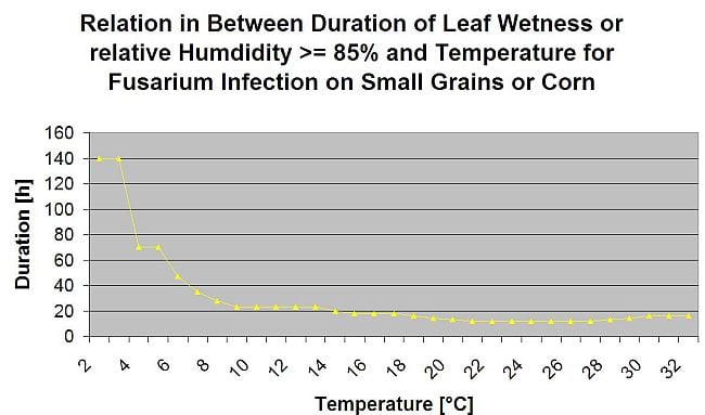


Summarizing all the different temperatures and moisture combinations we found in literature we descided to point out Fusarium Head bligh Infections if temperature and leaf wetness period or periods with more than 85% relative humdity are exceding the values visualised in the following graph.
Infections are started by a rain of 2 mm. A Fusarium Head Blight Infection can be assumed if the infection progress value reaches 100%. The calculation of this progress value follwos the relation in between the duration of moist conditions and the temperature displayed in the graph above.
This model is used to visualise the infection days and the climate conditions during climate. The growers knowledge about the development stage of the different wheat varieties gives the possibility to decide an application of a curative spray immidiately after infection.
Fusarium Mycotoxin Alert
Infection trials with extended leaf wetness periods of Fusarium head blight lead to high contents in mycotoxin. From this information an leaf wetness period of 48 hour or more during stage 61 and 69 is assumed to give a high risk of mycotoxins. Experiences made troughout the analyces of DON in commercial grown wheat showed that leaf wetness periods long enough for infection following an initial infection in stage 61 to 69 can increase the DON values too. In case of longer leaf wetness periods mycotoxins can increase up to stage 85. FieldClimate accumulates a risk figure proportional to the infection progress for every succesful infection periods over the period which has been selected to be propriate for this calculations. 6 just completed infection periods would lead to a risk of 100%. Normally the leaf wetness period leading to a fusarium infection is longer than in miniumum needed. Therefore most fusarium infections will lead to an risk increase of more than 17%. The risk value pointing to a problematic mycotoxin situation is depending on the field history. Wheat grown after non tillage corn or non tillage wheat after non tillage corn can only carry a small risk if it is not sprayed at the optimum situation. In unsprayed wheat we have to expect increased DON values already after 35% of risk. Wheat after non tillage wheat following any other crop than corn or wheat can carry a higher risk of 50%. If we have wheat after corn or wheat with tillage the risk being accsted can be extended to 70%. First year wheat shoulld be tested for DON if the risk exceeds 100%.
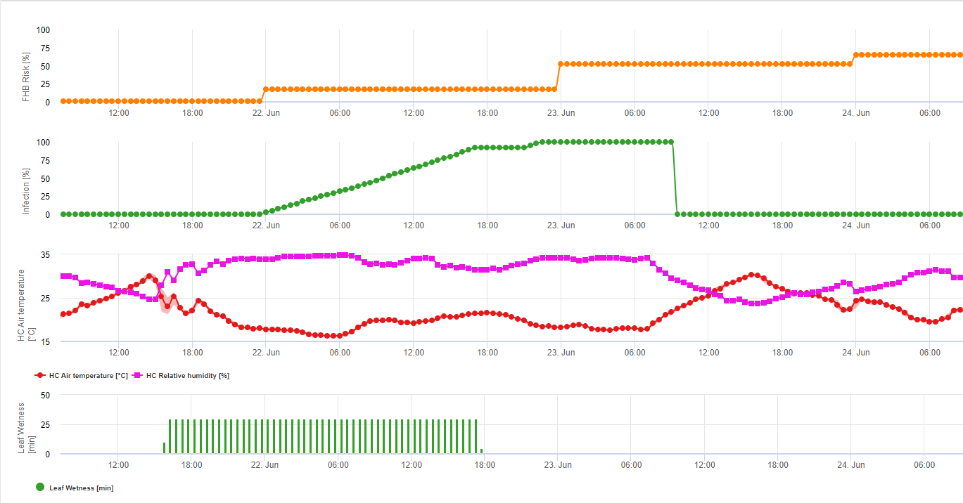
1) Fusarium Head Blight Risk model which figures out risky time periodes for an infection. Whenever 100% infection (green line) are reached the risk (blue line) is very high and conditions for the fungus have been favourable for infection. In dependance of the application method (curative, preventive) the risky time period is shown by the blue line.
2) Fusarium Head Blight: in this model the infection of the FHB is calculated by precipitation (2mm needed), relative humidity (above 85%) or leaf wetness, temperature during process. If infection reach 100% optimal conditions for the fungal pathogen have been reached. Further on the model figures out the FHB mycotoxin risk.
Speckled leaf blotch
There are two major Septoria diseases in wheat. These are Septoria tritici blotch, incited by the fungus Septoria tritici (teleomorph: Mycophaerella graminicola), and Septoria nodorum blotch, caused by the fungus Septoria nodorum (teleomorph: Leptosphaeria nodorum). Both diseases cause serious yield losses reported to range from 31 to 53 percent (Eyal, 1981; Babadoost and Herbert, 1984; Polley and Thomas, 1991). Worldwide, more than 50 million ha of wheat, mainly growing in the high-rainfall areas, are affected. During the past 25 years, these diseases have been increasing and have become a major limiting factor to wheat production in certain areas. Under severe epidemics, the kernels of susceptible wheat cultivars are shrivelled and are not fit for milling. Epidemics of Septoria tritici blotch and Septoria nodorum blotch of wheat are associated with favourable weather conditions (frequent rains and moderate temperatures), specific cultural practices, availability of inoculum and the presence of susceptible wheat cultivars (Eyal et al., 1987).
Septoria spp. Biology
Following Erick De Wolf, Septoria Tritici Blotch, Kansas State University, April 2008 Septoria tritici blotch known as speckled leaf blotch, is caused by the fungus Septoria tritici. It is distributed in all wheat-growing areas of the world and is a serious problem in many regions. Septoria tritici blotch is most damaging when the disease attacks the upper leaves and heads of susceptible varieties late in the season.
Symptoms
Septoria tritici blotch symptoms first appear in the fall. The initial symptoms are small yellow spots on the leaves. These lesions often become light tan as they age, and the fungal fruiting bodies can be seen embedded in the lesions on the awns. Lesions are irregularly shaped and range from elliptical to long and narrow (Figure 1). Lesions contain small, round, black speckles that are the fruiting bodies of the fungus. The black fruiting bodies look like grains of black pepper and can usually be seen without the aid of a magnifying glass. The disease begins on the lower leaves and gradually progresses to the flag leaf. Leaf sheaths are also susceptible to attack. In wet years, the speckled leaf blotch fungus can move onto the heads and cause brown lesions on the glumes and awns known as glume blotch. These lesions often become light tan as they age and the fungal fruiting bodies are often seen embedded in the lesions on the awns.
The glume blotch phase can cause significant yield loss, but the relationship between disease severity and yield loss is not well understood. Septoria tritici blotch can be confused with other leaf diseases that have very similar symptoms: tan spot and Stagonspora nodorum blotch, for example. It is common for plants to be infected by more than one of these foliar diseases, and it may require laboratory examination to accurately diagnose which diseases are most prevalent. Laboratory examination is nearly always required to distinguish the cause of glume blotch. Knowing the species is not important for spray decisions because all three diseases respond similarly to fungicides. However, knowing which diseases are most prevalent is an important part of variety selection because different genes control the resistance to the diseases.
The most reliable way to distinguish Septoria tritici blotch from the other diseases is by the presence of the black fungal fruiting bodies. The fungus that causes tan spot does not produce this type of reproductive structure. However, under moist conditions, the fungus that causes Stagonospora nodorum blotch will produce light brown fruiting bodies. In addition to the color difference, these structures are also smaller than those produced by Septoria tritici.
Life Cycle
Septoria tritici survives through the summer on residues of a previous wheat crop and initiates infections in the fall. There is some evidence that the fungus is able to survive in association with other grass hosts and wheat seed. These sources of the fungus are probably most important when the wheat residues are absent. Regardless of rotation or residue management practices, there is usually enough inoculum to initiate fall infections. Septoria tritici blotch is favored by cool, wet weather. The optimum temperature range is 16 to 21 °C; however, infections can occur during the winter months at temperatures as low as 5°C. Infection requires at least 6 hours of leaf wetness, and up to 48 hours of wetness are required for maximum infection. Once infection has occurred, the fungus takes 21 to 28 days to develop the characteristic black fruiting bodies and produce a new generation of spores. The spores produced in these fruiting bodies are exuded in sticky masses and require rain to splash them onto the upper leaves and heads.
Infection by Septoria tritici
Pycnidiospores of S. tritici germinate in free water from both ends of the spore or from intercalary cells (Weber, 1922). Spore germination does not begin until about 12 hours after contact with the leaf. Germ tubes grow randomly over the leaf surface. Weber (1922) observed only direct penetration between epidermal cells, but others concluded that penetration through both open and closed stomata is the primary means of host penetration (Benedict, 1971; Cohen and Eyal, 1993; Hilu and Bever, 1957). Kema et al. (1996) observed only stomatal penetration. Hyphae growing through stomata become constricted to about 1 μm diameter, then become wider after reaching the substomatal cavity.
Hyphae grow parallel to the leaf surface under epidermal cells, then through the mesophyll to cells of lower the epidermis, but not into the epidermis. No haustoria are formed and hyphal growth is limited by sclerenchyma cells around the vascular bundles, except when hyphae are very dense. Vascular bundles are not invaded. Hyphae grow intercellularly along cell walls through the mesophyll, branching at a septum or middle of a cell. No macroscopic symptoms appear for about 9 days except for an occasional dead cell, but mesophyll cells die rapidly after 11 days. Pycnidia develop in substomatal chambers. Hyphae seldom grow into host cells (Hilu and Bever, 1957; Kema et al, 1996; Weber, 1922).
Successful infection only occurs after at least 20 hours of high humidity. Only a few brown flecks developed if leaves remained wet for 5-10 hours after spore deposition (Holmes and Colhoun, 1974) or up to 24 hours (Kema et al., 1996). Host-parasite relations are the same on resistant or susceptible wheats. Spore germination on the leaf surface is the same regardless of susceptibility. The number of successful penetrations is about the same, but hyphal growth is faster in susceptible cultivars, resulting in more lesions. Hyphae extend 44 Session 2 — B.M. Cunfer beyond the necrotic area in all cultivars. A toxin may play a role in pathogenesis (Cohen and Eyal, 1993; Hilu and Bever, 1957). In contrast, colonization was greatly reduced on a resistant line (Kema et al., 1996).
Stagonospora (Septoria) and Septoria Pathogens of Cereals: The Infection Process
B.M. Cunfer, Department of Plant Pathology, University of Georgia, Griffin, GA
The infection process has been studied most intensely for Stagonospora (Septoria) nodorum and Septoria tritici. One in-depth study on Septoria passerinii is available. Nearly all of the information reported is for infection by pycnidiospores. However, the infection process for other spore forms is quite similar. The information presented is mostly for infection of leaves under optimum conditions. Some studies were done with intact seedling plants, whereas others were conducted with detached leaves. Infection of the wheat coleoptile and seedling by S. nodorum was described in detail by Baker (1971) and reviewed by Cunfer (1983). Although no precise comparisons have been made, it appears that the infection process has many similarities in each host-parasite system and is typical of many necrotrophic pathogens. Information on factors influencing symptom development and disease expression are excluded but have been reviewed by other authors (Eyal et al., 1987; King et al., 1983; Shipton et al., 1971). A summary of factors affecting spore longevity on the leaf surface is included.
Role of the Cirrus and Spore Survival on the Leaf Surface The most detailed information on the function of the cirrus encasing the pycnidiospores exuded from the pycnidium is for S. nodorum. The cirrus is a gel composed of proteinatous and saccharide compounds. Its composition and function are similar to that of other fungi in the Sphaeropsidales (Fournet, 1969; Fournet et al., 1970; Griffiths and Peverett, 1980). The primary roles of cirrus components are protection of pycnidiospores from dessication and prevention of premature germination.
The cirrus protects the pycnidiospores so that some remain viable at least 28 days (Fournet, 1969). When the cirrus was diluted with water, if the concentration of cirrus solution was >20%, less that 10% of pycnidiospores germinated. At a lower concentration, the components provide nutrients that stimulate spore germination and elongation of germ tubes. Germ tube length increased up to 15% cirrus concentration, then declined moderately at higher concentrations (Harrower, 1976). Brennan et al. (1986) reported greater germination in dilute cirrus fluid. Cirrus components reduced germination at 10-60% relative humidity. Once spores are dispersed, the stimulatory effects of the cirrus fluid are probably negligible (Griffiths and Peverett, 1980).
At 35-45% relative humidity, spores of S. tritici in cirri remained viable at least 60 days (Gough and Lee, 1985). The cirrus components may act as an inhibitor of spore germination, or the high osmotic potential of the cirrus may prevent germination. Pycnidiospores of S. nodorum did not survive for 24 hours at relative humidity above 80% at 20 C. Spores survived two weeks or more at <10% relative humidity (Griffiths and Peverett, 1980). When the cirrus fluid of S. nodorum was diluted with water, about two thirds of the pycnidiospores lost viability within 8 hours, and after 30 hours in daylight, only 5% germinated. When spores were stored in the dark, 40% remained viable after 30 hours (Brennan et al., 1986).
Dry conidia of S. nodorum, shaded and in direct sunlight, survived outdoors at least 56 hours (Fernandes and Hendrix, 1986a). Germination of S. nodorum pycnidiospores was inhibited by continuous UV-B (280-320 nm), whereas germination of S. tritici was not. Germ tube extension under continuous UV-B was inhibited for both fungi, compared with darkness (Rasanayagam et al., 1995).
Infection by Septoria nodorum
The process of host penetration and development of S. nodorum within the leaf was examined in detail by several investigators (Baker and Smith, 1978, Bird and Ride 1981, Karjalainen and Lounatmaa, 1986; Keon and Hargreaves, 1984; Straley, 1979; Weber, 1922). Pycnidiospores tend to lodge in the depressions between two epidermal cells, and many attempted leaf penetrations begin there. Spores germinate on the leaf surface in response to free moisture (Fernandes and Hendrix, 1986b). They begin to germinate 2-3 hours after deposition, and after 8 hours germination can reach 90%. Leaf penetration begins about 10 hours after spore deposition (Bird and Ride, 1981; Brönnimann et al., 1972; Holmes and Colhoun, 1974).
At the onset of germination, the germ tube is surrounded by an amorphous material that attaches to the leaf. Germ tubes growing from either end of a spore and from intercalary cells tend to grow along the depressions between cells and are often oriented along the long axis of the leaf (O’Reilly and Downes, 1986). Hyphae from spores not in depressions grow randomly with occasional branching (Straley, 1979). An appressorium forms with an infection peg that penetrates the cuticle and periclinal walls of epidermal cells directly into the cell lumen, resulting in rapid cell death.
Many penetrations first are subcuticular or lateral growth of a hypha occurs within the cell wall before growth into the cytoplasm (Bird and Ride, 1981; O’Reilly and Downes, 1986). Penetration through both open and closed stomata also occurs and may be faster than direct penetration (Harrower, 1976; Jenkins, 1978; O’Reilly and Downes, 1986; Straley, 1979). Germ tubes branch at stomata and junctions of epidermal cells. Penetration of a germ tube into a stomate may occur without formation of an appressorium. Penetration sometimes occurs through trichomes (Straley, 1979). Apparently, most penetration attempts fail, with dense papillae formed in the cells at the site of attempted penetration (Karjalainen and Lounatmaa, 1986; Bird and Ride, 1981).
After penetration, epidermal cells die quickly and become lignified, and the hyphae grow into the mesophyll. Mesophyll cells become misshapen, and lignified material is deposited outside of some cells, which then collapse. Lignification occurs before hyphae reach the cell. The process is the same in all cultivars but develops more slowly in resistant cultivars. The hyphae grow intercellularly among epidermal cells, then into the mesophyll. When the mesophyll is penetrated, chloroplast deterioration begins in 6-9 days (Karjalainen and Lounatmaa, 1986).
However, the photosynthetic rate begins to decline within a day after infection and before symptoms are visible (Krupinsky et al, 1973). Sclerenchyma tissue around vascular bundles prevents infection of vascular tissue. The vascular bundles block the spread of hyphae through the mesophyll except when sclerenchyma tissue is young and not fully formed (Baker and Smith, 1978).
Stagonospora nodorum releases a wide range of cell wall degrading enzymes including amylase, pectin methyl esterase, polygalacturonases, xylanases, and cellulase in vitro and during infection of wheat leaves (Baker, 1969; Lehtinen, 1993; Magro, 1984). The information related to cell wall degradation by enzymes agrees with histological observations.These enzymes may act in conjunction with toxins. Enzyme sensitivity may be related to resistance and rate of fungal colonization (Magro, 1984). Like many necrotrophs, Septoria and Stagonospora pathogens produce phytotoxic compounds in vitro. Cell deterioration and death in advance of hyphal growth into mesophyll tissue (Bird and Ride, 1981) is consistent with toxin production. However, a definitive role for toxins in the infection process and their relation to host resistance has not been established (Bethenod et al, 1982; Bousquet et al, 1980; Essad and Bousquet, 1981; King et al, 1983). Differences in host range between wheat and barleyadapted strains of S. nodorum may be related to toxin production (Bousquet and Kollmann, 1998). Initiation of spore germination and percentage of spores germinated are not influenced by host susceptibility (Bird and Ride, 1981; Morgan 1974; Straley, 1979; Straley and Scharen, 1979; Baker and Smith, 1978).
Bird and Ride (1981) reported that extension of germ tubes on the leaf surface was slower on resistant than on susceptible cultivars. This mechanism, expressed at least 48 hours after spore deposition, indicates pre-penetration resistance to elongation of germ tubes. There were fewer successful penetrations in resistant cultivars, and penetration proceeded more slowly on resistant cultivars (Baker and Smith, 1978; Bird and Ride, 1981). Lignification was proposed to limit infection in both resistant and susceptible cultivars, but other factors slowed fungal development in resistant lines. In susceptible lines, faster growing Hyphae may escape lignification of host cells.Four days after inoculation of barley with a wheat biotype isolate of S. nodorum, hyphae grew through the cuticle and sometimes in outer cellulose layers of epidermal cell walls. Thick papillae were deposited beneath the penetration hyphae and the cells were not penetrated (Keon and Hargreaves, 1984).
Infection by Septoria passerinii
Green and Dickson (1957) present a detailed description of the infection process of S. passerinii on barley. The infection process is similar to S. tritici. Like S. tritici, the length of time required for leaf penetration is considerably longer than for S. nodorum. Germ tubes branch and grow over the leaf surface at random, but sometimes along depressions between epidermal cells. Leaf penetration is almost exclusively through stomata. Germination hyphae become swollen, and if penetration is unsuccessful, hyphae continue to elongate. No penetration occurs 48 hours after spore deposition. After 72 hours, germ tubes thicken over stomata, grow between guard cells and on urfaces of accessary cells and into the substomatal cavities. Direct penetration between epidermal cells is seen only rarely.
Spore germination and host penetration are the same on resistant and susceptible cultivars. There is much less extension of hyphae within leaves on resistant cultivars and papillae are observed on many but not all cell walls. Hyphae grow beneath the epidermis from one stoma to another, but do not penetrate between epidermal cells. The mesophyll is colonized, but no haustoria form. After the mesophyll cells become necrotic, epidermal cells collapse. Mycelial development in the leaf is sparse and usually blocked by vascular bundles. In younger leaves, if the vascular sheath is less developed, hyphae pass between the bundle and the epidermis. Pycnidia form in substomatal cavities, mostly on the upper leaf surface (Green and Dickson, 1957).
Factors Affecting Spore Longevity on the Leaf Surface Among the Stagonospora and Septoria pathogens of cereals, definitive information on the infection process has been reported only for S. nodorum, S. tritici, and S. passerinii. Like many other necrotrophic pathogens, neither group of pathogens elicit the hypersensitive reaction. A significant difference in the infection process between Septoria and Stagonospora pathogens is that spore germination and penetration proceeds much faster for S. nodorum than for S. tritici and S. passerinii. This has a significant influence on disease epidemiology.
The Septoria pathogens penetrate the plant primarily through stomata, whereas S. nodorum penetrates both directly and through stomata. S. nodorum penetrates and kills the epidermal cells quickly, but S. tritici and S. passerinii do not kill epidermal cells until hyphae have ramified through the leaf mesophyll and rapid necrosis begins. Histological studies of fungal growth following host penetration match the data generated from epidemiological studies of host resistance. Resistance slows the rate of host colonization but has no appreciable effect on the process of lesion development.
The mechanisms controlling host response, whether related to enzymes and toxins or other metabolites released by the pathogens during infection, are still unclear. There is little information about infection by ascospores. The infection process is probably very similar to that for pycnidiospores. Ascospores of Phaeosphaeria nodorum germinate over a wide range of temperatures, and their germ tubes penetrate the leaf directly. However, according to Rapilly et al. (1973), ascospores, unlike pycnidiospores, do not germinate in free water.
Septoria spp. Infection Model
Septoria Infections are possible at low temperatures whereas temperatures below 7°C might not lead to an infections within 2 days. The optimum temperature of the disease is reached in the area of 16 to 21°C. Infections are possible within a period of high relative humditiy or leaf wetness of 14 hours or longer. To meet the conditions we decided to seperate into models for weak, moderate and severe infections. Weak infections can be given if it is possible for the pathogen to infect the host tissue. This means that weak infections can take place if temperatures are in miniumum and leaf wetness periods are of critical duration. A moderate infection will take place under conditions where most infeciton trials lead to reasonable results and severe infections take place under conditions where the pathogen has optimum conditions for infection.
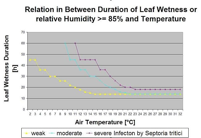


Infection starts after a rain of 0.5 mm. We decided not to use a model for pycnidia formation. The condition needed for pycnidia formation is assumed to be a period with relative humdity higher than 85%. Pycnidia life time is expected to be 24 hours. In all climates where Septoria tritici has a chance to infect we will find 2 hours fulfilling this conditions at nearly every day arround sun rise.
Infection severity evaluation
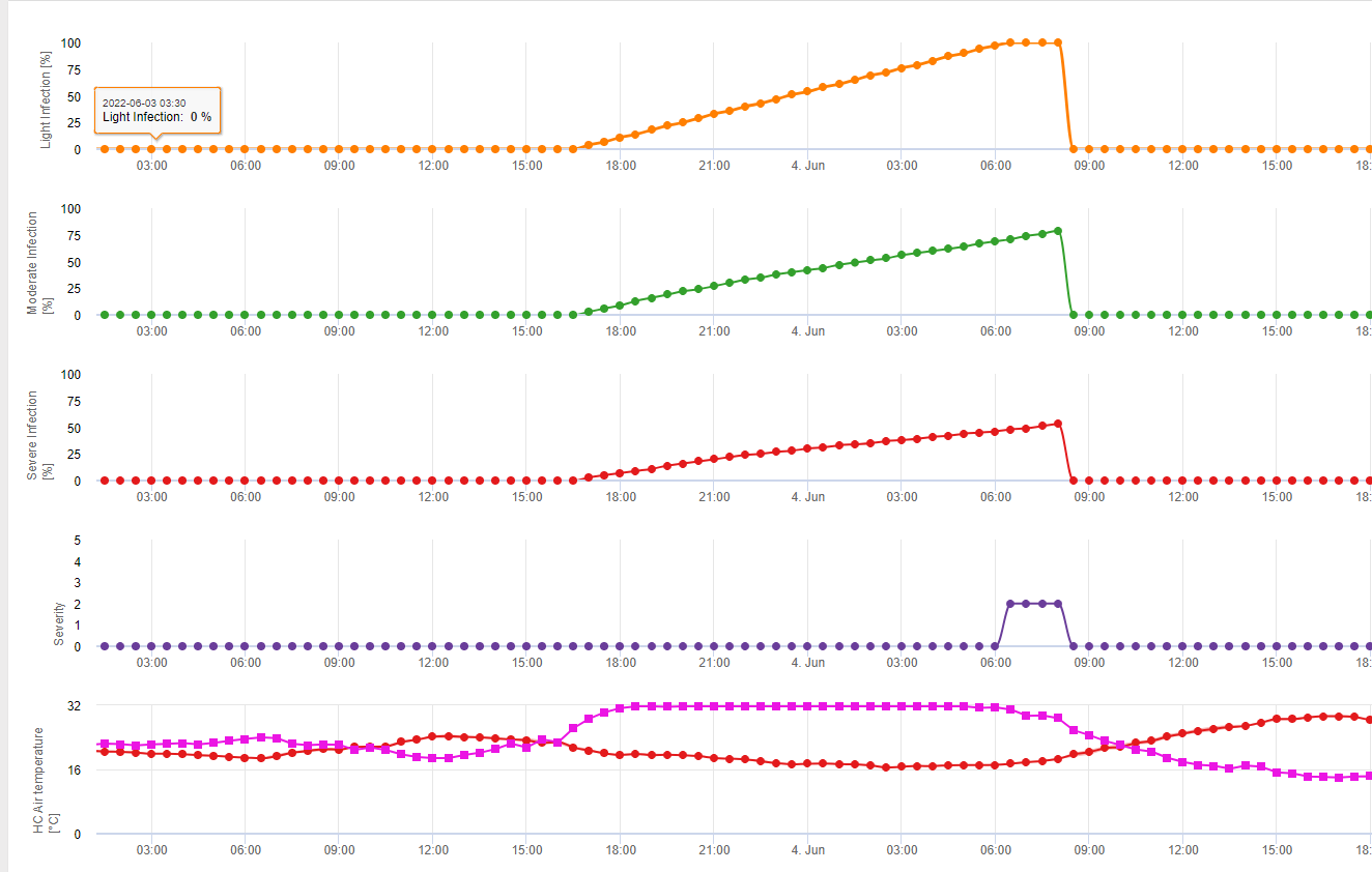
To be able to assess the Septoria tritici infection pressure in between stage 10 (first leaf trough coleoptile) and stage 32 (node two at least 2 cm above node 1) and in between 32 and 51 (beginning of heading) we have to assess the severity of infections based on climatic conditions. This assesment is done in a 1 to 5 scale. A severity of 1 is given if the condition for a weak infection is fullfiled and it has rained less than 5 mm, otherwise the corresponding sevirity vlaue will be 2. A severity of 3 is given if a moderate infection is fullfilled and it has rained less than 5mm. If it has rained more than 5 mm during a moderate infection or less than 5 mm during a severe infection a severity of 4 is given.
A severe infection with more than 5 mm of rain corresponds with a severity value of 5.
Septoria tritici disease pressure evaluation
The climate is only one factor desciding about the disease pressure in field. The other two factors are the history of the field and the susceptibility of the variety grown. If we can accumulate the disease severity values from stage 10 to stage 32 to value of 4 we can expect a weak disease pressure by the climate. If this value reaches 6 we can expect a moderate disease pressure and if it reaches 10 we can expect a high disease pressure from the climate. Knowing the susceptibility of the variety and the history of the field will lead us to spray or not on a weak or moderate disease pressure in this situation. Having an accumulated value of 10 may lead to a spray in stage 32 anyhow. Decision of a spray at a later stage is more depending on the spring climate. If we are able to accumulate the severity values since stage 10 to a value of 6 we can expect a weak disease pressure. If this value reaches 10 we can expect an moderate disease pressure and if this value reaches 15 we can expect a high disease pressure form the climate situation.
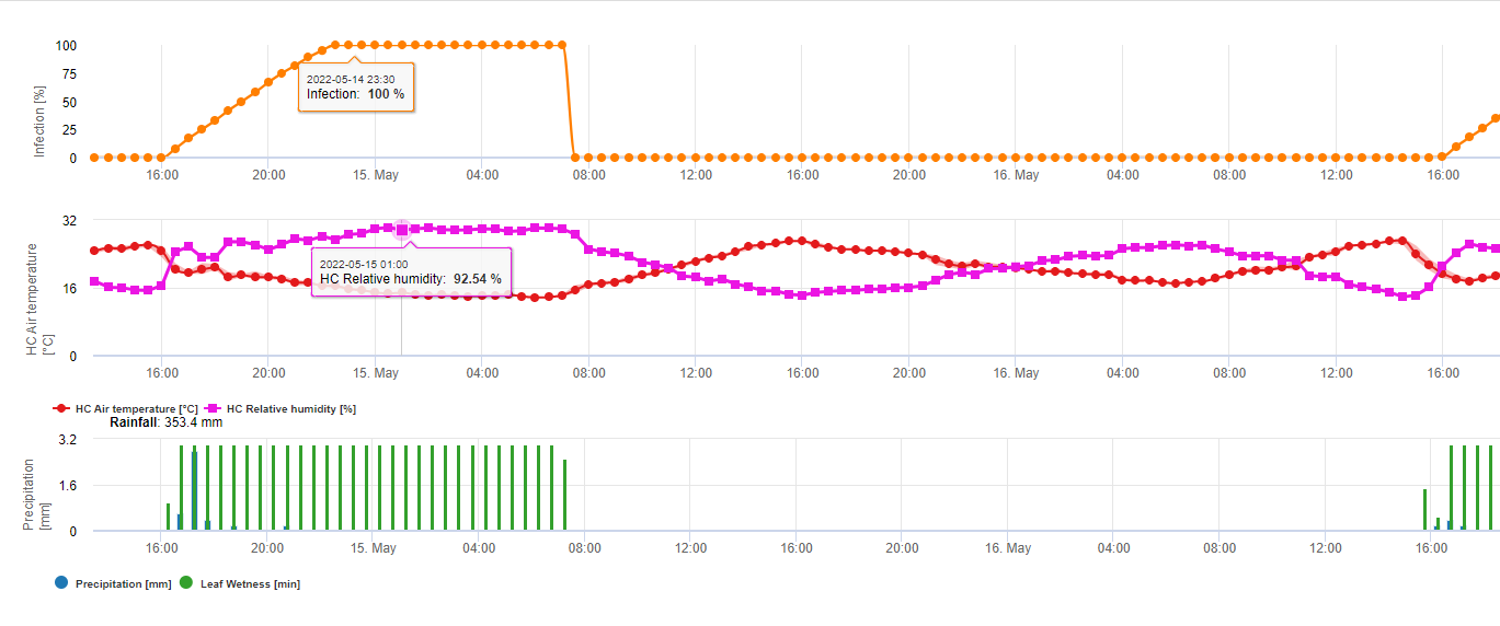
In FieldClimate we show the Septoria tritici Severity together with the three different infection severities in one graph (see above). Due too rainfall and long leaf wetness periodes conditions for a severe infection by S. tritici have been fullfield on May 14th and 16th. The Severity levels reach the highest value of 5 on May 14th, which means that a high risk for infection is now.
Stagonospora nodorum’s infection biology differs in some extend form this of S. tritici but this difference is not big enough for a seperate model. Therfore we suggest to use this model for the whole complex of Stagnospora and Septoria diseases in cereals including S. passerinii. S. tritici and S. passerinii tend to need longer leaf wetness periods than S. nodorum. In areas with a high pressure of S. nodorum infections classifed to a weak giving a severity value of 2 should be treated more serious then in other areas.
For Septoria nodorum a risk model is shown in FieldClimate (see above). A high risk was determined at the June 17th and July 7th (100%). Depending on the susceptible plant stage for infection plant protection measurements have to bee taken into account if the risk reach 80% (also see wheather forecast, protective plant protection). If the risk was 100% and an infection was already determined systemic plant protection measurements (curative application) has to be taken to protect the plant.
Rice blast
In the tropics, blast spores are present in the air throughout the year, thus favoring continuous development of the disease. The infection brought about by the fungus damages upland rice severely than the irrigated rice. It rarely attacks the leaf sheaths. Primary infection starts where seed is sown densely in seedling boxes for mechanical transplanting.
In the temperate countries, it overseasons in infested crop residue or in seed. Cloudy skies, frequent rain, and drizzles favor the development and severity of rice blast. High nitrogen levels, high relative humidity, and wet leaves encourage infection caused by the fungus. The rate of sporulation is highest with increasing relative humidity of 90% or higher. For leaf wetness, the optimum temperature for germination of the pathogen is 25-28 °C. Growing rice in aerobic soil in wetlands where drought stress is prevalent also favors infection.
A fungus causes rice blast. Its conidiophores are produced in clusters from each stoma. They are rarely solitary with 2-4 septa. The basal area of the conidiophores is swollen and tapers toward the lighter apex. The conidia of the fungus measure 20-22 x 10-12 µm. The conidia are 2-septate, translucent, and slightly darkened. They are obclavate and tapering at the apex. They are truncate or extended into a short tooth at the base.
Besides the rice plant, the fungus also survives on Agropyron repens (L.) Gould, Agrostis palustris, A. tenuis, Alopecurus pratensis, Andropogon sp., Anthoxanthum odoratum, Arundo donax L., Avena byzantina, A. sterilis, A. sativa, Brachiaria mutica (Forssk.) Stapf, Bromus catharticus, B. inermis, B. sitchensis, Canna indica, Chikushichloa aquatica, Costus speciosus, Curcuma aromatica, Cynodon dactylon (L.) Pers., Cyperus rotundus L., C. compressus L., Dactylis glomerata, Digitaria sanguinalis (L.) Scop, Echinochloa crus-galli (L.) P. Beauv., Eleusine indica (L.) Gaertn., Eragrostis sp., Eremochloa ophiuroides, Eriochloa villosa, Festuca altaica, F. arundinacea, F. elatior, F. rubra, Fluminea sp., Glyceria leptolepis, Hierochloe odorata, Holcus lanatus, Hordeum vulgare, Hystrix patula, Leersia hexandra Sw., L. japonica, L. oryzoides, Lolium italicum, L. multiflorum, L. perenne, Muhlenbergia sp., Musa sapientum, Oplismenus undulatifolius (Ard.) Roem. & Schult., Panicum miliaceum L., P. ramosum (L.) Stapf, P. repens L., Pennisetum typhoides (L.) R. Br., Phalaris arundinacea L., P. canariensis, Phleum pratense, Poa annua L., P. trivialis, Saccharum officinarum, Secale cereale, Setaria italica (L.) P. Beauv., S. viridis (L.) P. Beauv., Sorghum vulgare, Stenotaphrum secundatum, Triticum aestivum, Zea mays L., Zingiber mioga, Z. officinale, and Zizania latifolia.
Conidia are produced on lesions on the rice plant about 6 days after inoculation. The production of spores increases with increase in the relative humidity. Most of the spores are produced and released during the night. After spore germination, infection follows. Infection tubes are formed from the appressoria and later the penetration through the cuticle and epidermis. After entering the cell, the infection tube forms a vesicle to give rise to hyphae. In the cell, the hyphae grew freely.
Rice blast infects the rice plant at any growth stage. Rice seedlings or plants at the tillering stage are often completely killed. Likewise, heavy infections on the panicles usually cause a loss in rice yields.
Anthracnose
The Model of R. solani in fields is evaluating the risk for this disease on base of temperature, leaf wetness and global radiation. It checks last 120 hours:
- In case of consecutive leaf wetness it accumulate temperature depending values for every minute.
– 12 °C to 15 °C it accumulates 1 per minute
– 16 °C to 17 °C it accumulates 2 per minute
– 18°C and more it accumulates 4 per minute - At the end of the leaf wetness periods it evaluates the accumulated values
– if they are greater than 4096 it increases risk by 64 points and subtracts 4096 from the value>
– if the residual is greater than 2048 it increases the risk by 16 and subtracts 2048 from the value
– if the residual is greater than 1024 it increases the risk by 4 and subtracts 1024 form the value - If global radiation is consecutive higher than 800 W/m² accumulate time in minutes and if radiation becomes lower evaluate values:
– Value > 512 = RiskValue – 32 Points , Value – 512
– Value > 256 = RiskValue – 8 Points , Value – 256
– Value >128= RisKValue – 2 Points, Value -128
The model results in a risk value between 0 and 100 indicating the times favorable for R. solani in fields.
Support System
The model points out periods with a high risk for this disease. No sprays will have to be applied in periods where the risk is low. In periods with moderate risk spray interval can be prolongated and in periods with high risk spray interval may have to be reduced or more effective compounds will have to be used.
Literature
Rust Diseases
- Anikster, Y., Bushnell, W.R., Eilam, T., Manisterski, J. & Roelfs, A.P. 1997. Puccinia recondita causing leaf rust on cultivated wheats, wild wheats, and rye. Can. J. Bot., 75: 2082-2096.
- Azbukina, Z. 1980. Economical importance of aecial hosts of rust fungi of cereals in the Soviet Far East. In Proc. 5th European and Mediterranean Cereal Rusts Conf., p.199-201. Bari, Rome.
- Beresford, R.M. 1982. Stripe rust (Puccinia striiformis), a new disease of wheat in New Zealand. Cer. Rusts Bull., 10: 35-41.
- Biffen, R.H. 1905. Mendel’s laws of inheritance and wheat breeding. J. Agric. Sci., 1: 4-48.
- Borlaug, N.E. 1954. Mexican wheat production and its role in the epidemiology of stem rust in North America. Phytopathology, 44: 398-404.
- Buchenauer, H. 1982. Chemical and biological control of cereal rust. In K.J. Scott & A.K. Chakravorty, eds. The rust fungi, p. 247-279. London, Academic Press.
- Burdon, J.J., Marshall, D.R. & Luig, N.H. 1981. Isozyme analysis indicates that a virulent cereal rust pathogen is somatic hybrid. Nature, 293: 565-566.
- Casulli, F. 1988. Overseasoning of wheat leaf rust in southern Italy. In Proc. 7th European and Mediterranean Cereal Rusts Conf., 5-9 Sept., p. 166-168. Vienna.
- Chester, K.S. 1946. The nature and prevention of the cereal rusts as exemplified in the leaf rust of wheat. In Chronica botanica. Walthan, MA, USA. 269 pp.
- Craigie, J.H. 1927. Experiments on sex in rust fungi. Nature, 120: 116-117.
- Cummins, G.B. & Caldwell, R.M. 1956. The validity of binomials in the leaf rust fungus complex of cereals and grasses. Phytopathology, 46: 81-82.
- Cummins, G.B. & Stevenson, J.A. 1956. A check list of North American rust fungi (Uredinales). Plant Dis. Rep. Suppl., 240: 109-193.
- d’Oliveira, B. & Samborski, D.J. 1966. Aecial stage of Puccinia recondita on Ranunculaceae and Boraginaceae in Portugal. In Proc. Cereal Rusts Conf., Cambridge, p 133-150.
- DeBary, A. 1866. Neue Untersuchungen uber die Uredineen insbesondere die Entwicklung der Puccinia graminis und den Zusammenhang duselben mit Aecidium berberis. In Monatsber Preuss. Akad. Wiss., Berlin, p. 15-20.
- de Candolle, A. 1815. Uredo rouille des cereales. In Flora francaise, famille des champignons, p. 83.
- Dubin, H.J. & Stubbs, R.W. 1986. Epidemic spread of barley stripe rust in South America. Plant Dis., 70: 141-144.
- Dubin, H.J. & Torres, E. 1981. Causes and consequences of the 1976-1977 wheat leaf rust epidemic in northwest Mexico. Ann. Rev. Phytopath., 19: 41-49.
- Eriksson, J. 1894. Uber die Spezialisierung des Parasitismus bei dem Getreiderostpilzen. Ber. Deut. Bot. Ges., 12: 292-331.
- Eriksson, J. & Henning, E. 1894. Die Hauptresultate einer neuen Untersuchung uber die Getreideroste. Z. Pflanzenkr., 4: 66-73, 140-142, 197-203, 257-262.
- Eriksson, J. & Henning, E. 1896. Die Getreideroste. Ihre Geschichte und Natur sowie Massregein gegen dieselben, p. 463. Stockholm, P.A. Norstedt and Soner.
- Ezzahiri, B., Diouri, S. & Roelfs, A.P. 1992. The-Role of the alternate host, Anchusa italica, in the epidemiology of Puccinia recondita f. sp. tritici on durum wheats in Morocco. In F.J. Zeller & G. Fischbeck, eds. Proc. 8th European and Mediterranean Cereal Rusts and Mildews Conf., 1992, p. 69-70. Heft 24 Weihenstephan, Germany.
- Fontana, F. 1932. Observations on the rust of grain. P.P. Pirone, transl. Classics No. 2. Washington, DC, Amer. Phytopathol. Society. (Originally published in 1767).
- Green, G.J. & Campbell, A.B. 1979. Wheat cultivars resistant to Puccinia graminis tritici in western Canada: their development, performance, and economic value. Can. J. Plant Pathol., 1: 3-11.
Groth, J.V. & Roelfs, A.P. 1982. The effect of sexual and asexual reproduction on race abundance in cereal rust fungus populations. Phytopathology, 72: 1503-1507. - Hare, R.A. & McIntosh, R.A. 1979. Genetic and cytogenetic studies of the durable adult plant resistance in ‘Hope’ and related cultivars to wheat rusts. Z. Pflanzenzüchtg, 83: 350-367.
- Hart, H. & Becker, H. 1939. Beitrage zur Frage des Zwischenwirts fur Puccinia glumarum Z. Pflanzenkr. (Pflanzenpathol.) Pflanzenschutz, 49: 559-566.
- Hendrix, J.W., Burleigh, J.R. & Tu, J.C. 1965. Oversummering of stripe rust at high elevations in the Pacific Northwest-1963. Plant Dis. Rep., 49: 275-278.
- Hermansen, J.E. 1968. Studies on the spread and survival of cereal rust and mildew diseases in Denmark. Contrib. No. 87. Copenhagen, Department of Plant Pathology, Royal Vet. Agriculture College. 206 pp.
- Hirst, J.M. & Hurst, G.W. 1967. Long-distance spore transport. In P.H. Gregory & J.L. Monteith, eds. Airborne microbes, p. 307-344. Cambridge University Press.
- Hogg, W.H., Hounam, C.E., Mallik, A.K. & Zadoks, J.C. 1969. Meteorological factors affecting the epidemiology of wheat rusts. WMO Tech. Note No. 99. 143 pp.
- Huerta-Espino, J. & Roelfs, A.P. 1989. Physiological specialization on leaf rust on durum wheat. Phytopathology, 79: 1218 (abstr.).
- Hylander, N., Jorstad, I. & Nannfeldt, J.A. 1953. Enumeratio uredionearum Scandinavicarum. Opera Bot., 1: 1-102.
- Johnson, R. 1981. Durable disease resistance. In J.F. Jenkyn & R.T. Plumb, eds. Strategies for control of cereal diseases, p. 55-63. Oxford, UK, Blackwell.
- Johnson, T. 1949. Intervarietal crosses in Puccinia graminis. Can. J. Res. Sect. C, 27: 45-63.
- Joshi, L.M. & Palmer, L.T. 1973. Epidemiology of stem, leaf, and stripe rusts of wheat in northern India. Plant Dis. Rep., 57: 8-12.
- Katsuya, K. & Green, G.J. 1967. Reproductive potentials of races 15B and 56 of wheat stem rust. Can. J. Bot., 45: 1077-1091.
- Kilpatrick, R.A. 1975. New wheat cultivars and longevity of rust resistance, 1971-1975. USDA, Agric. Res. Serv. Northeast Reg (Rep.), ARS-NE 64. 20 pp.
- Le Roux, J. & Rijkenberg, F.H.J. 1987. Pathotypes of Puccinia graminis f. sp. tritici with increased virulence for Sr24. Plant Dis., 71: 1115-1119.
- Lewellen, R.T., Sharp, E.L. & Hehn, E.R. 1967. Major and minor genes in wheat for resistance to Puccinia striiformis and their response to temperature changes. Can. J. Bot., 45: 2155-2172.
- Line, R.F. 1976. Factors contributing to an epidemic of stripe rust on wheat in the Sacramento Valley of California in 1974. Plant Dis. Rep., 60: 312-316.
- Line, R.F., Qayoum, A. & Milus, E. 1983. Durable resistance to stripe rust of wheat. In Proc. 4th Int. Cong. Plant Pathol., p. 204. Melbourne.
- Luig, N.H. 1985. Epidemiology in Australia and New Zealand. In A.P. Roelfs & W.R. Bushnell, eds. Cereal rusts, vol. 2, Diseases, distribution, epidemiology, and control, p. 301-328. Orlando, FL, USA, Academic Press.
- Luig, N.H. & Rajaram, S. 1972. The effect of temperature and genetic background on host gene expression and interaction to Puccinia graminis tritici. Phytopathology, 62: 1171-1174.
- Luig, N.H. & Watson, I.A. 1972. The role of wild and cultivated grasses in the hybridization of formae speciales of Puccinia graminis. Aust. J. Biol. Sci., 25: 335-342.
- Maddison, A.C. & Manners, J.G. 1972. Sunlight and viability of cereal rust urediospores. Trans. Br. Mycol. Soc., 59: 429-443.
- Mains, E.B. 1932. Host specialization in the leaf rust of grasses Puccinia rubigovera. Mich. Acad. Sci., 17: 289-394.
- Mains, E.B. 1933. Studies concerning heteroecious rust. Mycologia, 25: 407-417.
- Mains, E.B. & Jackson, H.S. 1926. Physiologic specialization in the leaf rust of wheat, Puccinia triticina Erikss. Phytopathology, 16: 89-120.
- Manners, J.G. 1960. Puccinia striiformis Westend. var. dactylidis var. nov. Trans. Br. Mycol. Soc., 43: 65-68.
- McIntosh, R.A. 1976. Genetics of wheat and wheat rusts since Farrer: Farrer memorial oration 1976. J. Aust. Inst. Agric. Sci., 42: 203-216.
- McIntosh, R.A. 1992. Close genetic linkage of genes conferring adult-plant resistance to leaf rust and stripe rust in wheat. Plant Pathol., 41: 523-527.
- McIntosh, R.A., Luig, N.H., Milne, D.L. & Cusick, J. 1983. Vulnerability of triticales to wheat stem rust. Can. J. Plant Pathol., 5: 61-69.
- McIntosh, R.A., Hart, G.E., Devos, K.M., Gale, M.D. & Rogers, W.J. 1998. Catalogue of gene symbols for wheat. In Proc. 9th Int. Wheat Genetics Symp., 2-7 Aug. 1998. Saskatoon Saskatchewan, Canada.
- Melander, L.W. & Craigie, J.H. 1927. Nature of resistance of Berberis spp. to Puccinia graminis. Phytopathology, 17: 95-114.
- Mont, R.M. 1970. Studies of nonspecific resistance to stem rust in spring wheat. M.Sc. thesis. St Paul, MN, USA, University of Minnesota. 61 pp.
- Nagarajan, S. & Joshi, L.M. 1985. Epidemiology in the Indian subcontinent. In A.P. Roelfs & W.R. Bushnell, eds. The cereal rusts, vol. 2, Diseases, distribution, epidemiology, and control, p. 371-402. Orlando, FL, USA, academic Press.
- O’Brien, L., Brown, J.S., Young, R.M. & Pascoe, T. 1980. Occurrence and distribution of wheat stripe rust in Victoria and susceptibility of commercial wheat cultivars. Aust. Plant Pathol. Soc. Newsl., 9: 14.
- Perez, B. & Roelfs, A.P. 1989. Resistance to wheat leaf rust of land cultivars and their derivatives. Phytopathology, 79: 1183 (abstr.).
- Pretorius, Z.A., Singh, R.P., Wagoire, W.W. & Payne, T.S. 2000. Detection of virulence to wheat stem rust resistance gene Sr31 in Puccinia graminis f. sp. tritici in Uganda. Phytopathology, 84: 203.
- Rajaram, S., Singh, R.P. & van Ginkel, M. 1996. Approaches to breed wheat for wide adaptation, rust resistance and drought. In R.A. Richards, C.W. Wrigley, H.M. Rawson, G.J. Rebetzke, J.L. Davidson & R.I.S. Brettell, eds. Proc. 8th Assembly Wheat Breeding Society of Australia, p. 2-30. Canberra, The Australian National University.
- Rapilly, F. 1979. Yellow rust epidemiology. Ann. Rev. Phytopathol., 17: 5973.
- Robbelen, G. & Sharp, E.L. 1978. Mode of inheritance, interaction, and application of genes conditioning resistance to yellow rust. Fortschr. Pflanzenzucht. Beih. Z. Pflanzenzucht., 9: 1-88.
- Roelfs, A.P. 1978. Estimated losses caused by rust in small grain cereals in the United States 1918-1976. Misc. Publ. USDA, 1363: 1-85.
Roelfs, A.P. 1982. Effects of barberry eradication on stem rust in the United States. Plant Dis., 66: 177-181. - Roelfs, A.P. 1985a. Epidemiology in North America. In A.P. Roelfs & W.R. Bushnell, eds. The cereal rusts, vol. 2, Diseases, distribution, epidemiology, and control, p. 403-434. Orlando, FL, USA, Academic Press.
- Roelfs, A.P. 1985b. Wheat and rye stem rust. In A.P. Roelfs & W.R. Bushnell, eds. The cereal rusts, vol. 2, Diseases, distribution, epidemiology, and control, p. 3-37. Orlando, FL, USA, Academic Press.
- Roelfs, A.P. 1986. Development and impact of regional cereal rust epidemics. In K.J. Leonard & W.E. Fry, eds. Plant disease epidemiology, vol. 1, p. 129-150. New York, NY, USA, MacMillan.
- Roelfs, A.P. 1988. Genetic control of phenotypes in wheat stem rust. Ann. Rev. Phytopathol., 26: 351-367.
- Roelfs, A.P. & Groth, J.V. 1980. A comparison of virulence phenotypes in wheat stem rust populations reproducing sexually and asexually. Phytopathology, 70: 855-862.
- Roelfs, A.P. & Groth, J.V. 1988. Puccinia graminis f. sp. tritici, black stem rust of Triticum spp. In G.S. Sidhu, ed. Advances in plant pathology, vol. 6, Genetics of pathogenic fungi, p. 345-361. London, Academic Press.
- Roelfs, A.P. & Long, D.L. 1987. Puccinia graminis development in North America during 1986. Plant Dis., 71: 1089-1093.
- Roelfs, A.P. & Martell, L.B. 1984. Ure-dospore dispersal from a point source within a wheat canopy. Phytopathology, 74: 1262-1267.
- Roelfs, A.P., Huerta-Espino, J. & Marshall, D. 1992. Barley stripe rust in Texas. Plant Dis., 76: 538.
- Rowell, J.B. 1981. Relation of postpenetration events in Idaed 59 wheat seedling to low receptivity to infection by Puccinia graminis f. sp. tritici. Phytopathology, 71: 732-736.
- Rowell, J.B. 1982. Control of wheat stem rust by low receptivity to infection conditioned by a single dominant gene. Phytopathology, 72: 297-299.
- Rowell, J.B. 1984. Controlled infection by Puccinia graminis f. sp. tritici under artificial conditions. In. A.P. Roelfs & W.R. Bushnell, eds. The cereal rusts, vol. 1, Origins, specificity, structure, and physiology, p. 291-332. Orlando, FL, USA, Academic Press.
- Saari, E.E. & Prescott, J.M. 1985. World distribution in relation to economic losses. In A.P. Roelfs & W.R. Bushnell, eds. The cereal rusts, vol. 2, Diseases, distribution, epidemiology, and control, p. 259-298. Orlando, FL, USA, Academic Press.
Saari, E.E., Young, H.C., Jr. & Fernkamp, M.F. 1968. Infection of North American Thalictrum spp. with Puccinia recondita f. sp. tritici. Phytopathology, 58: 939-943. - Samborski, D.J. 1985. Wheat leaf rust. In A.P. Roelfs and W.R. Bushnell, eds. The cereal rusts, vol. 2, Diseases, distribution, epidemiology, and control, p. 39-59. Orlando, FL, USA, Academic Press.
- Savile, D.B.O. 1984. Taxonomy of the cereal rust fungi. In W.R. Bushnell & A.P. Roelfs, eds. The cereal rusts, vol. 1, Origins, specificity, structures, and physiology, p. 79-112. Orlando, FL, USA, Academic Press.
- Sharp, E.L. & Volin, R.B. 1970. Additive genes in wheat conditioning resistance to stripe rust. Phytopathology, 601: 1146-1147.
- Singh, R.P. 1992. Genetic association of leaf rust resistance gene Lr34 with adult plant resistance to stripe rust in bread wheat. Phytopathology, 82: 835-838.
- Singh, R.P. & Rajaram, S. 1992. Genetics of adult-plant resistance to leaf rust in “Frontana” and three CIMMYT wheats. Genome, 35: 24-31.
- Singh, R.P. & Rajaram, S. 1994. Genetics of adult plant resistance to stripe rust in ten spring bread wheats. Euphytica, 72: 1-7.
- Singh, R.P., Mujeeb-Kazi, A. & Huerta-Espino, J. 1998. Lr46: A gene conferring slow-rusting resistance to leaf rust in wheat. Phytopathology, 88: 890-894.
- Skovmand, B., Fox, P.N. & Villareal, R.L. 1984. Triticale in commercial agriculture: progress and promise. Adv. Agron., 37: 1-45.
- Spehar, V. 1975. Epidemiology of wheat rust in Europe. In Proc. 2nd Int. Winter Wheat Conf., 9-19 Jun., p. 435-440. Zagreb, Yugoslavia.
- Stakman, E.C. 1923. Wheat diseases. 14. The wheat rust problem in the United States. In Proc. 1st Pan Pac. Sci. Congr., p. 88-96.
- Straib, W. 1937. Untersuchungen uben dasn Vorkommen physiologischen Rassen der Gelbrostes (Puccinia glumarum) in den Jahren 1935-1936 und uber die Agressivitat eninger neuer Formes auf Getreide un Grasern. Arb. Biol. Reichsant. Land. Fortw. Berlin-Dahlem, 22: 91-119.
- Stubbs, R.W. 1985. Stripe rust. In A.P. Roelfs & W.R. Bushnell, eds. The cereal rusts, vol. 2, Diseases, distribution, epidemiology, and control, p. 61-101. Orlando, FL, USA, Academic Press.
- Stubbs, R.W. & de Bruin, T. 1970. Bestrijding van gele roest met het systemische fungicide oxycaroxin ‘Plantvax’. Gewasbescherming, 1: 99-104.
- Stubbs, R.W., Prescott, J.M., Saari, E.E. & Dubin, H.J. 1986. Cereal disease methodology manual, p. 46. Mexico, DF, CIMMYT.
- Sunderwirth, S.D. & Roelfs, A.P. 1980. Greenhouse evaluation of the adult plant resistance of Sr2 to wheat stem rust. Phytopathology, 70: 634-637.
- Tollenaar, H. & Houston, B.R. 1967. A study on the epidemiology of stripe rust, Puccinia striiformis West in California. Can. J. Bot., 45: 291-307.
- Tozzetti, G.T. 1952. V. Alimurgia: True nature, causes and sad effects of the rusts, the bunts, the smuts, and other maladies of wheat and oats in the field. In L.R. Tehon, transl. Phytopathological Classics No. 9, p. 139. St Paul, MN, USA, American Phytopathol. Society. (Originally published 1767).
- Tranzschel, W. 1934. Promezutocnye chozjaeva rzavwiny chlebov i ich der USSR. (The alternate hosts of cereal rust fungi and their distribution in the USSR). In Bull. Plant Prot., Ser. 2, p. 4-40. (In Russian with German summary).
- Viennot-Bourgin, G. 1934. La rouille jaune der graminees. Ann. Ec. Natl. Agric. Grignon Ser., 3, 2: 129-217.
- Wallwork, H. & Johnson, R. 1984. Transgressive separation for resistance to yellow rust in wheat. Euphytica, 33: 123-132.
- Watson, I.A. & de Sousa, C.N.A. 1983. Long distance transport of spores of Puccinia graminis tritici in the Southern Hemisphere. In Proc. Linn. Soc. N.S.W., 106: 311-321.
- Wright, R.G. & Lennard, J.H. 1980. Origin of a new race of Puccinia striiformis. Trans. Br. Mycol. Soc., 74: 283-287.
- Zadoks, J.C. 1961. Yellow rust on wheat studies of epidemiology and physiologic specialization. Neth. J. Plant Pathol., 67: 69-256.
- Zadoks, J.C. & Bouwman, J.J. 1985. Epidemiology in Europe. In A.P. Roelfs & W.R. Bushnell, eds. The cereal rusts , vol. 2, Diseases, distribution, epidemiology, and control , p. 329-369. Orlando, FL, USA, Academic Press
Fusarium Head Blight
- Baht, C.V., Beedu, R., Ramakrishna, Y. & Munshi, R.L. 1989. Outbreak of trichothecene toxicosis associated with consumption of mould-damaged wheat products in Kashmir Valley, India. Lancet, p. 35-37.
- Bai, G. 1995. Scab of wheat: epidemiology, inheritance of resistance and molecular markers linked to cultivar resistance. Ph.D. thesis. West Lafayette, IN, USA, Purdue University.
- Chen, P., Liu, D. & Sun, W. 1997. New countermeasures of breeding wheat for scab resistance. In H.J. Dubin, L. Gilchrist, J. Reeves & A. McNab, eds. Fusarium head scab: global status and future prospects. Mexico, DF, CIMMYT.
- Diaz de Ackermann, M. & Kohli, M.M. 1997. Research on fusarium head blight of wheat in Uruguay. In H.J. Dubin, L. Gilchrist, J. Reeves & A. McNab, eds. Fusarium head scab: global status and future prospects. Mexico, DF, CIMMYT.
- Dill-Macky, R. 1997. Fusarium head blight: Recent epidemics and research efforts in the upper midwest of United States. In H.J. Dubin, L. Gilchrist, J. Reeves & A. McNab, eds. Fusarium head scab: global status and future prospects. Mexico, DF, CIMMYT.
- Dubin, H.J., Gilchrist, L., Reeves, J. & McNab?, A. 1997. Fusarium head scab: global status and future prospects. Mexico, DF, CIMMYT.
- Galich, M.T.V. 1997. Fusarium head blight in Argentina. In H.J. Dubin, L. Gilchrist, J. Reeves & A. McNab, eds. Fusarium head scab: global status and future prospects. Mexico, DF, CIMMYT.
- Gilchrist, L., Rajaram, S., Mujeeb-Kazi?, A., van Ginkel, M.,Vivar, H. & Pfeiffer, W. 1997. In H.J. Dubin, L. Gilchrist, J. Reeves & A. McNab, eds. Fusarium head scab: global status and future prospects. Mexico, DF, CIMMYT.
- Ireta, J. 1989. Seleccion de un modelo de crecimiento para la fusariosis del trigo causada por Fusarium graminearum Schw. In M.M. Kohli, ed. Taller sobre la Fusariosis de la espiga en America del Sur. Mexico, DF, CIMMYT.
- Ireta, M.J. & Gilchrist, L. 1994. Fusarium head scab of wheat (Fusarium graminearum Schwabe). Wheat Special Report No. 21b. Mexico, DF, CIMMYT.
- Khonga, E.B. & Sutton, J.C. 1988. Inoculum production and survival of Giberella zeae in maize and wheat residues. Can. J. Plant Pathol., 10(3): 232-240.
- Luo, X.Y. 1988. Fusarium toxin contamination in China. Proc. Japan Assoc. Mycotoxicology Suppl., 1: 97-98. Marasas, W.F.O., Jaskiewickz, K., Venter, F.S. & Schalkwyk, D.J. 1988. Fusarium moniliforme contamination of maize in oesophageal cancer areas in Transkei. S. AF. Med. J., 74: 110-114.
- Mihuta-Grimm, L. & Foster, R.L. 1989. Scab on wheat and barley in southern Idaho and evaluation of seed treatment for eradication of Fusarium spp. Plant Dis., 73: 769-771.
- Miller, J.D. & Armison, P.G. 1986. Degradation of deoxynivalenol by suspension cultures of the Fusarium head blight resistant wheat cultivar Frontana. Can. J. Plant Pathol., 8: 147-150.
- Miller, J.D., Young, J.C. & Sampson, D.R. 1985. Deoxynivalenol and Fusarium head blight resistance in spring cereals. J. Phytopathol., 113: 359-367.
- Reis, E.M. 1985. Doencas do trigo III. Fusariose. Brasil, Merck Sharp Dohme.
- Reis, E.M. 1989. Fusariosis: biologia y epidemiologia de Gibberella zeae en trigo. In M.M. Kohli, ed. Taller sobre la Fusariosis de la espiga en America del Sur. Mexico, DF, CIMMYT.
- Schroeder, H.W. & Christensen, J.J. 1963. Factors affecting resistance of wheat to scab caused by Gibberella zeae. Phytopathology, 53: 831-838.
- Snidjers, C.H.A. 1989. Current status of breeding wheat for Fusarium head blight resistant and mycotoxin problem in the Netherlands. Foundation of Agricultural Plant Breeding, Wageningen. In M.M. Kohli, ed. Taller sobre la Fusariosis de la espiga en America del Sur. Mexico, DF, CIMMYT.
- Snijders, C.H.A. 1990. The inheritance of resistance to head blight caused by Fusarium culmorum in winter wheat Euphytica, 50: 11-18.
- Teich, A.H. & Nelson, K. 1984. Survey of Fusarium head blight and possible effects of cultural practices in wheats fields in Lambton County in 1983. Can. Plant Dis. Surv., 64(1): 11-13.
- Trenholm, H.L., Friend, D.H. & Hamilton, R.M.G. 1984. Vomitoxin and zearalenone in animal feeds. Agriculture Canada Publication 1745 E.
- Van Egmond, H.P. & Dekker, W.H. 1995. Worldwide regulations for mycotoxins in 1994. Nat. Toxins, 3: 332.
- Viedma, L. 1989. Importancia y distribucion de la fusariosis de trigo en Paraguay. In M.M. Kohli, ed. Taller sobre la Fusariosis de la espiga en America del Sur. Mexico, DF, CIMMYT.
- Wang, Y.Z. 1997. Epidemiology and management of wheat scab in China In H.J. Dubin, L. Gilchrist, J. Reeves & A. McNab, eds. Fusarium head scab: global status and future prospects. Mexico, DF, CIMMYT.
- Wang, Y.Z. & Miller, J.D. 1988. Effects of metabolites on wheat tissue in relation to Fusarium head blight resistance. J. Phytopathol., 122: 118-125.
Fusarium Head Blight
- Infektion und Ausbreitung von Fusarium spp. an Weizen in Abhängigkeit der Anbaubedingungen im Rheinland, Lienemann, K.
- E.-C. Oerke und H.-W. Dehne
Water activity, temperature, and pH effects on growth of Fusarium moniliforme and Fusarium proliferatum isolates from maize, Sonia Marin, Vicente Sanchis, and Naresh Magan - Interaction of Fusarium graminearum and F. moniliforme in Maize Ears: Disease Progress, Fungal Biomass, and Mycotoxin Accumulation, L. M. Reid, R. W. Nicol, T. Ouellet, M. Savard, J. D. Miller, J. C. Young, D. W. Stewart, and A. W. Schaafsma
- Evaluierung von Einflussfaktoren auf den Fusarium-Ährenbefall des Weizens, Wolf, P. F. J.; Schempp, H.; Verreet, J.-A.
Septoria spp.
- Baker, C.J. 1971. Morphology of seedling infection by Leptosphaeria nodorum. Trans. Brit. Mycol. Soc. 56:306-309.
- Baker, C.J. 1969. Studies on Leptosphaeria nodorum Müller and Septoria tritici Desm. on wheat. Ph. D. thesis. University of Exeter.
- Baker E.A., and Smith, I.M. 1978. Development of the resistant and susceptible reactions in wheat on inoculation with Septoria nodorum. Trans. Brit. Mycol. Soc. 71:475-482.
- Benedict, W.G. 1971. Differential effect of light intensity on the infection of wheat by Septoria tritici Desm. under controlled environmental conditions. Physiol. Pl. Pathol. 1:55-56.
- Bird, P.M., and Ride, J.P. 1981. The resistance of wheat to Septoria nodorum: fungal development in relation to host lignification. Physiol. Pl. Pathol. 19:289-299.
- Stagonospora and Septoria Pathogens of Cereals: The Infection Process 45 Bethenod, O., Bousquet, J., Laffray, D., and Louguet, P. 1982. Reexamen des modalités d’action de l’ochracine sur la conductance stomatique des feuilles de plantules de blé, Triticum aestivum L., cv “Etoile de Choisy”.Agronomie 2:99-102.
- Bousquet, J.F., and Kollman, A. 1998. Variation in metabolite production by Septoria nodorum isolates adapted to wheat or to barley. J. Phytopathol. 146:273-277.
- Bousquet, J.F., Belhomme de Franqueville, H., Kollmann, A., and Fritz, R. 1980. Action de la septorine, phytotoxine synthétisée par Septoria nodorum, sur la phosphorylation oxydative dans les mitochondries isolées de coléoptiles de blé. Can. J. Bot. 58:2575-2580.
- Brennan, R.M., Fitt, B.D.L., Colhoun, J., and Taylor, G.S. 1986. Factors affecting the germination of Septoria nodorum pycnidiospores. J. Phytopathol. 117:49-53.
- Brönnimann, A., Sally, B.K., and Sharp, E.L. 1972. Investigations on Septoria nodorum in spring wheat in Montana. Plant Dis. Reptr. 56:188-191.
- Cohen, L., and Eyal, Z. 1993. The histology of processes associated with the infection of resistant and susceptible wheat cultivars with Septoria tritici. Plant Pathol. 42:737-743.
- Cunfer, B.M. 1983. Epidemiology and control of seed-borne Septoria nodorum. Seed Sci. Tecnol. 11:707- 718.
- Essad, S., and Bousquet, J.F. 1981. Action de l’ochracine, phytotoxine de Septoria nodorum Berk., sur le cycle mitotique de Triticum aestivum L. Agronomie 1:689-694.
- Eyal, Z., Scharen, A.L., Prescott, J.M., and van Ginkel, M. 1987. The Septoria diseases of wheat: Concepts and methods of disease management. CIMMYT. Mexico D.F. 46 pp.
- Fernandes, J.M.C., and Hendrix, J.W. 1986a. Viability of conidia of Septoria nodorum exposed to natural conditions in Washington. Fitopathol. Bras. 11:705-710.
- Fernandes, J.M.C., and Hendrix, J.W. 1986b. Leaf wetness and temperature effects on the hyphal growth and symptoms development on wheat leaves infected by Septoria nodorum. Fitopathol. Bras. 11:835-845.
- Fournet, J. 1969. Properiétés et role du cirrhe du Septoria nodorum Berk. Ann. Phytopathol. 1:87-94. Fournet, J., Pauvert, P., and Rapilly, F. 1970. Propriétés des gelées sporifères de quelques Sphaeropsidales et Melanconiales. Ann. Phytopathol. 2:31-41.
- Gough, F.J., and Lee, T.S. 1985. Moisture effects on the discharge and survival of conidia of Septoria tritici. Phytopathology 75:180-182.
- Green, G.J., and Dickson, J.G. 1957. Pathological histology and varietal reactions in Septoria leaf blotch of barley. Phytopathology 47:73-79.
- Griffiths, E., and Peverett, H. 1980. Effects of humidity and cirrhus extract on survival of Septoria nodorum spores. Trans. Brit. Mycol. Soc. 75:147-150.
- Harrower, K.M. 1976. Cirrus function in Leptosphaeria nodorum. APPS Newsletter 5:20-21.
- Hilu, H.M., and Bever, W.M. 1957. Inoculation, oversummering, and suscept-pathogen relationship of on Triticum species. Phytopathology 47:474-480.
- Holmes, S.J.I., and Colhoun, J. 1974. Infection of wheat by Septoria nodorum and S. tritici. Trans. Brit. Mycol. Soc. 63:329-338.
- Jenkins, P.D. 1978. Interaction in cereal diseases. Ph. D. Thesis. University of Wales.
- Karjalainen, R., and Lounatmaa, K. 1986. Ultrastructure of penetration and colonization of wheat leaves by Septoria nodorum. Physiol. Mol. Pl. Pathol. 29:263-270.
- Kema, G.H.J., DaZhao, Y., Rijkenberg, F.H.J., Shaw, M.W., and Baayen, R.P. 1996. Histology of the pathogenesis of Mycosphaerella graminicola in wheat. Phytopathology 86:777-786.
- Keon, J.P.R., and Hargreaves, J.A. 1984. The response of barley leaf epidermal cells to infection by Septoria nodorum. New Phytol. 98:387-398.
- King, J.E., Cook, R.J., and Melville, S.C. 1983. A review of Septoriadiseases of wheat and barley. Ann.Appl. Biol. 103:345-373.
- Krupinsky, J.M., Scharen, A.L., and Schillinger, J.A. 1973. Pathogen variation in Septoria nodorum Berk., in relation to organ specificity, apparent photosynthetic rate and yield of wheat. Physiol. Pl. Pathol. 3:187-194.
- Lehtinen, U. 1993. Plant cell wall degrading enzymes of Septoria nodorum. Physiol. Mol. Pl. Pathol. 43:121-134.
- Magro, P. 1984. Production of polysacchride-degrading enzymes in culture and during pathogenesis. Plant Sci. Letters 37:63-68.
- Morgan, W.M. 1974. Physiological studies of diseases of wheat caused by Septoria spp. and Fusarium culmorum. Ph. D. thesis. University of London.
- O’Reilly, P., and Downes, M.J. 1986. Form of survival of Septoria nodorum on symptomless winter wheat. Trans. Brit. Mycol. Soc. 86:381-385.
- Rapilly, F., Foucault, B., and Lacazedieux, J. 1973. Études sur l’inoculum de Septoria nodorum Berk. (Leptosphaeria nodorum Müller) agent de la septoriose du blé I. Les ascospores. Ann. Phytopathol. 5:131-141.
- Rasanayagam, M.S., Paul, N.D., Royle, D.J., and Ayres, P.G. 1995. Variation in responses of Septoria tritici and S. nodorum to UV-B irradiation in vitro. Mycol. Res. 99:1371-1377.
- Shipton, W.A., Boyd, W.R.J., Rosielle, A.A., and Shearer, B.L. 1971. The common Septoria diseases of wheat. Bot. Rev. 37:231-262.
- Straley, M. L. 1979. Pathogenesis of Septoria nodorum (Berk.) Berk. On wheat cultivars varying in resistance to glume blotch. Ph. D. thesis. Montana State University. 99 pp.
- Straley, M.L., and Scharen, A.L. 1979. Development of Septoria nodorum in resistant and susceptible wheat leaves. Phytopathology 69:920-921.
- Weber, G.F. 1922. Septoria diseases of cereals. Phytopathology 12:537-585.
- Babadoost, M. & Herbert, T.T. 1984. Factors affecting infection of wheat seedlings by Septoria nodorum. Phytopathology, 74: 592-595.
- Eyal, Z.E. 1981. Integrated control of Septoria diseases of wheat. Plant Dis., 65: 763-768.
- Eyal, Z., Sharen, A.L., Prescott, J.M. & van Ginkel, M. 1987. The Septoria diseases of wheat: concepts and methods of disease management. Mexico, DF, CIMMYT.
- Polley, R.W. & Thomas, M.R. 1991. Surveys of disease of wheat in England and Wales, 1976-1988. Ann. Appl. Biol., 119: 1-20.
- Henze, Mathias: Entwicklung einer anbauparameter- und witterunsabhängigen Befallsprognose von Septoria tritici, CUVILLER VERLAG, Göttingen 2008
- B.M. Cunfer: Stagonospora and Septoria Pathogens of Cereals: The Infection Process, in Septoria and Stagonospora Diseases of Cereals: A Compilation of Global Research, M. van Ginkel, A. McNab, and J. Krupinsky editors, Proceedings of the Fifth International Septoria Workshop September 20-24, 1999, CIMMYT, Mexico
Recommended equipment
Check which sensor set is needed for monitoring this crop’s potential diseases.

